


 下载:
下载:

-
成像光谱仪是一种非常高效的定量探测仪器,它可以获得待观测目标或场景的连续单色光谱图像,并通过空间维(x, y)数据和光谱维(λ)数据共同组成三维观测数据立方体,从而为研究人员提供探测目标或场景中的每一个点的空间和光谱特征细节[1-3]。这种兼具相机的高分辨成像能力和光谱仪的高光谱分辨能力使得成像光谱仪被广泛应用于测绘遥感、目标识别、环境监测与评估、临床影像诊断、过程监控等任务中,并在地理、空间、海洋、气候、大气、农业、植被、生态、医药、安防、制造与色度等多个领域中发挥巨大作用[4-8]。
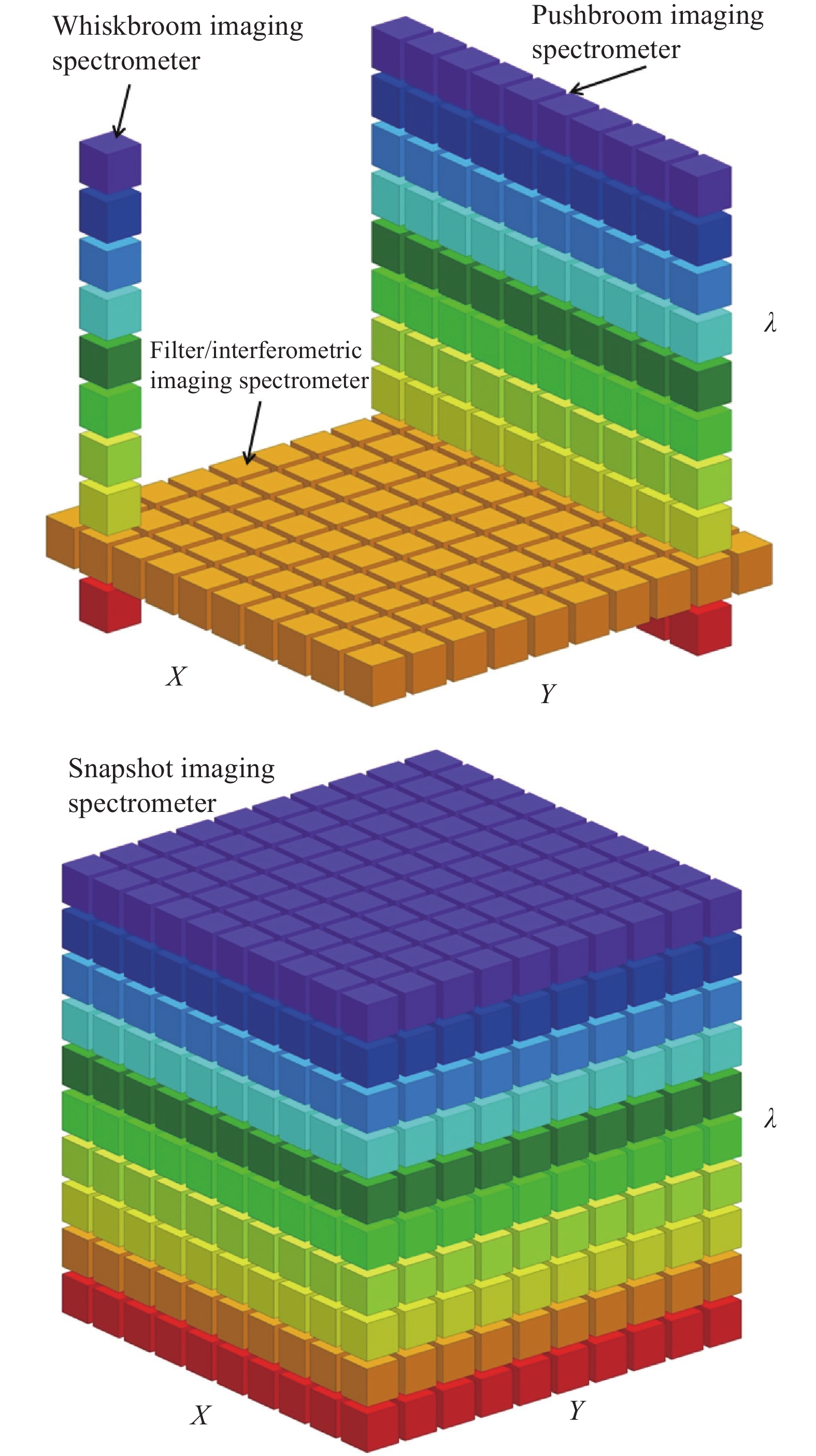
成像光谱仪概念的提出已较难考证,最早提出概念的时间大约在20世纪20~30年代。1945年,Beutler首次系统地给出了凹面光栅的光程数学分析理论[9];1967年,Werner又系统地阐述了衍射光栅的几何光学像差理论[10]。这些理论为成像光谱仪的建立和发展奠定了基础。随着现代光学的快速发展,成像光谱仪也得到了长足的进步,各种类型层出不穷,分光器件形式五花八门,性能上也有了很大的提高。但归根结底,所有成像光谱仪的最终探测目的都是为了获取目标场景的三维立方数据体,故根据其获取数据过程的不同可以将成像光谱仪进行最基本的分类,如图1所示:
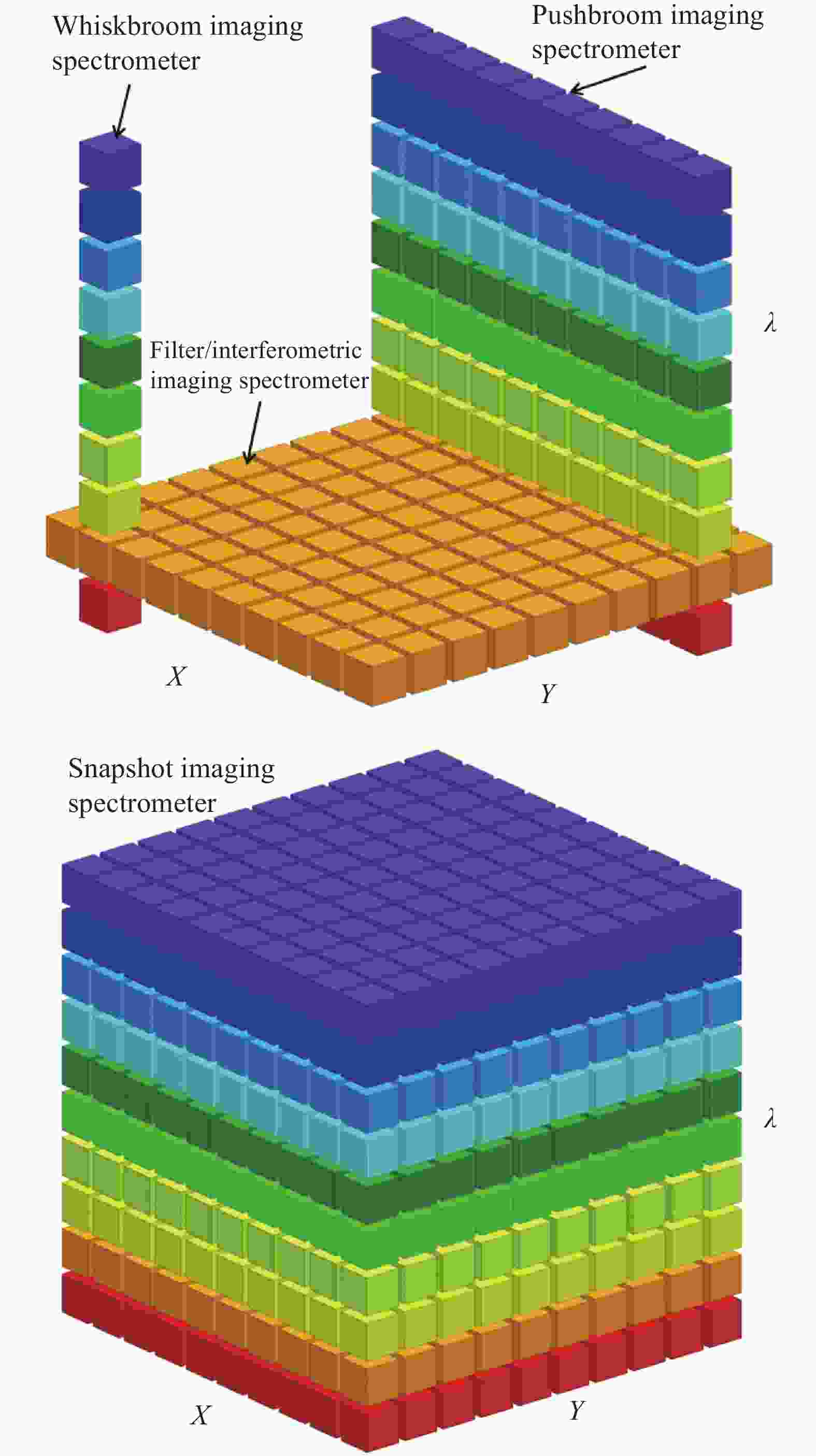
图 1 不同成像光谱仪在单次观测积分时间下获取三维体立方数据过程示意图
Figure 1. Portions of the datacube collected during a single observing integration period for different imaging spectrometers
(1) 摆扫式成像光谱仪(Whiskbroom imaging spectrometer),使用线阵探测器,每次收集一个像素上的所有光谱维数据,并通过扫描镜摆动和搭载平台的移动最终获取全部的立方数据体,相对来说获取速度最慢。
(2) 推扫式成像光谱仪(Pushbroom imaging spectrometer),使用面阵探测器,每次可以收集一维空间维上对应的一行像素的所有光谱维数据,并通过搭载平台的移动或扫描镜的转动完成全部数据立方体的收集。
(3) 滤光片式成像光谱仪(Filter/imaging spectrometer),使用面阵探测器,通过一组滤光片或可调谐波长器件进行分光,每次可以收集两个空间维度上的面阵对应的所有像素的单一光谱维数据,并通过滤光片切换或器件的波长调谐来完成全部数据立方体收集。干涉型成像光谱仪(如:傅里叶变换光谱仪)等均可归入此类。
(4) 快照型成像光谱仪(Snapshot imaging spectrometer),使用面阵探测器,通过特殊元器件或探测形式对目标或场景进行编码,再通过后续系统色散后对探测器采集数据进行解码,一次观测即可收集整个三维数据立方体,探测速度最快;但是其光谱分辨率不高,且数据的解码速度较慢。
一般而言,成像光谱仪系统主要包括了前置成像系统、光谱色散成像系统和探测器系统,其中用于激发光谱观测的仪器在前置成像系统前可能还会放置激发光源。成像光谱仪的核心部分是光谱色散成像系统,该系统决定了成像光谱仪的主要性能参数,也是仪器在进行设计和研制时所需完成的最关键部分。因此,文中将根据成像光谱仪中的光谱色散成像系统获取光谱信息能力的不同,按照滤光型成像光谱仪、色散型成像光谱仪、干涉型成像光谱仪和快照型成像光谱仪的分类对各类光谱仪的原理、主要光学形式和应用进行介绍。
-
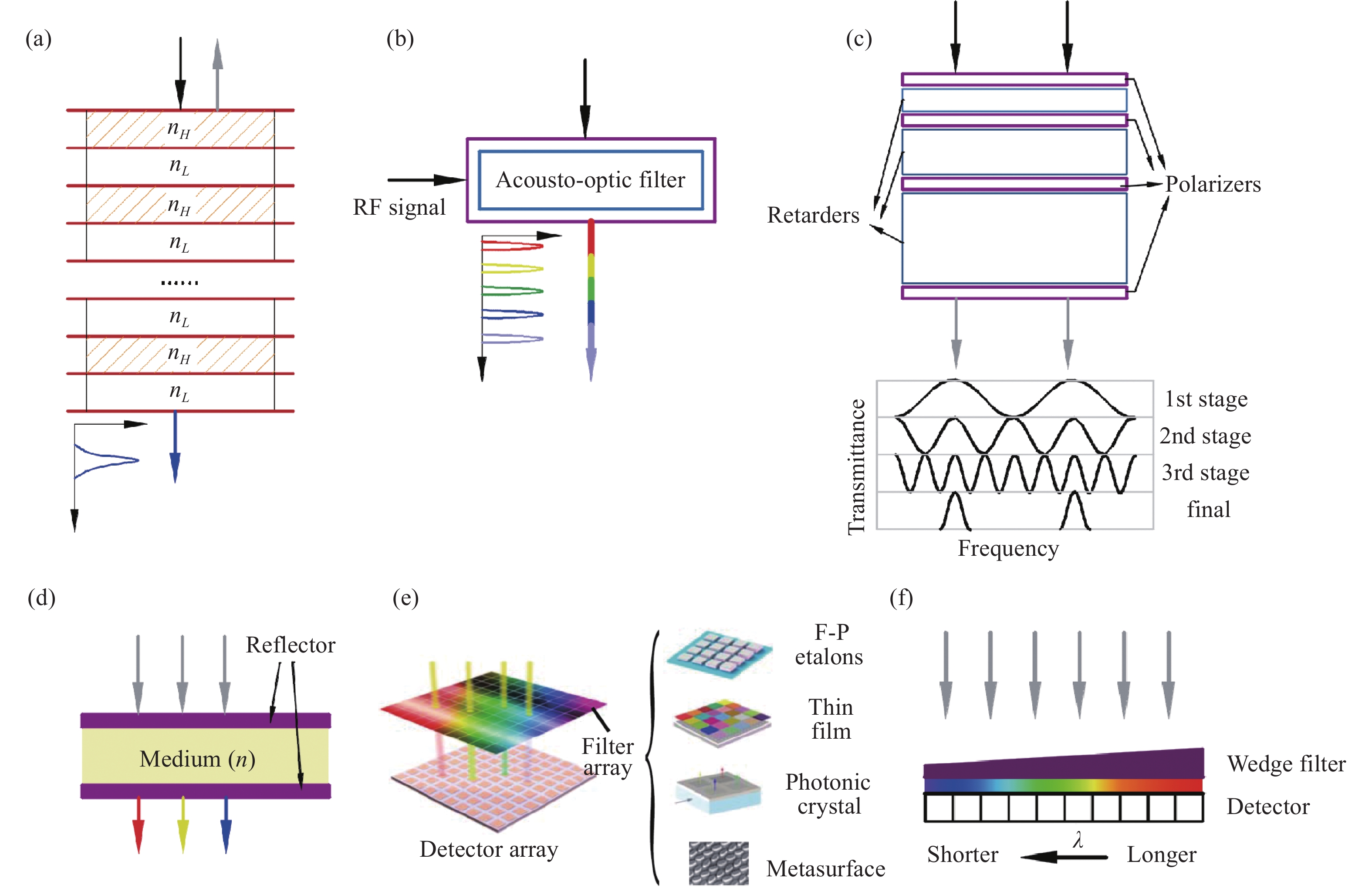
窄带滤光片是一种能够对探测目标辐射光中的特征光谱进行有效提取、同时对带外杂光进行高抑制的光学器件,将窄带滤光片置于相机中的探测器前即可实现相机对拟观测目标的特征光谱图像探测,从而形成最简单形式的成像光谱仪[11]。如果观测目标或场景具备多个特征波长,则可以使用多个窄带滤光片组成的滤光片组,通过分时切换来实现多波长的单色光谱成像探测。
这种成像光谱仪的核心光谱色散器件和系统形式相比其他成像光谱仪而言较小,因此整体结构上更容易实现集成化和小型化。目前,很多市面上使用的可在无人机上搭载的小型高光谱相机均属于此类成像光谱仪,其体积小、质量轻、成本低,在农业定量观测等领域中应用较为广泛。
一般的窄带滤光片带宽在几纳米到十几甚至几十纳米之间,但有时为了实现极窄带宽的特征光谱观测,就需要使用干涉型滤光片。这种滤光片的制作一般需要使用多层膜技术,通过在滤光片玻璃基底上交替镀一系列具有不同折射率的膜层材料,入射光将在薄膜各层界面处经过反复的反射和折射,从而最终在出射端获得极窄的光谱谱线(原理见图2(a))。这种极窄线宽的干涉型滤光片相对价格比较昂贵,多用于天文观测和实验室定量测试的成像光谱仪器中。

图 2 滤光型成像光谱仪主要分光原理:(a) 干涉式滤光片;(b) 声光可调谐滤光器;(c) Lyot-Ohman液晶双折射可调谐滤光器;(d) 法布里-珀罗腔;(e) 阵列式滤光器; (f) 线性渐变式滤光片
Figure 2. Main principle of filter imaging spectrometers: (a) Interference filter;(b) Acousto-optic tunable filter;(c) Lyot-Ohman liquid crystal birefingent tunable filter;(d) Fabry-Perot etalon;(e) Array filter; (F) Linear variable filter
-
目前主流的可调谐窄带滤光器包括声光可调谐滤光器(AOTF)[12]、液晶双折射可调谐滤光器(LCTF)[13]、法布里-珀罗(F-P)滤光器[14]等。下面对这几种滤光器做简要介绍。
-
这种滤光器主要利用声光衍射原理,器件的核心是晶体和其相连的压电换能器,通过将高频的射频电信号作用于换能器,使之转换为在晶体内的超声波振动,这种振动波可以在传播区域内周期性地调制晶体折射率,从而产生空间周期性的调制作用,这种调制与体相位光栅类似,可以对入射到晶体介质上的电磁波进行衍射,而且衍射光的波长与高频驱动电信号的频率一一对应,只要改变高频RF驱动信号的频率即可改变衍射光的波长,达到分光的目的,如图2(b)所示。
声光可调谐滤光器可动态地选择特定波长,并以微秒级的速度进行波长调整。
-
以液晶双折射可调谐滤光器中最典型的Lyot-Ohman滤光器为例进行说明(原理如图2(c)所示)。这种滤光器由液晶延迟波片和线性偏振片按照一定的顺序组合而成。所有的延迟波片其快慢轴的指向均一致,且与其相邻的偏振片轴成π/4夹角;且一般而言每个延迟波片的厚度都是前一个的2倍。令φ为晶体中产生的相位差,则其传输函数可表示为[15]:
$$ T = {\cos ^2}\left( {\frac{\varphi }{2}} \right) = {\cos ^2}\left( {\frac{{\pi \Delta }}{\lambda }} \right) = {\cos ^2}\left( {\frac{{\pi {{d}}\delta n}}{\lambda }} \right) $$ (1) 式中:∆为延迟量;δn为沿晶体轴线的折射率微量差值;d为晶体厚度,λ为波长。这样,一个完整的Lyot-Ohman滤光器的传输函数就可以表示为:
$$ T={T}_{1}\cdot{T}_{2}\cdot{T}_{3}\cdots{T}_{n}=\left(\frac{\mathrm{sin}\left({2}^{N}\varphi /2\right)}{{2}^{N}\mathrm{sin}\left(\varphi /2\right)}\right) $$ (2) 与声光可调谐滤光器工作原理类似,通过对液晶双折射滤光器中的晶体施加电场,可产生电控双折射现象,从而改变o光和e光的折射率差,进而通过电压精确调制二者的相位差来实现不同波长的输出。Lyot-Ohman滤光器的一大缺点是偏振片对入射光辐射的吸收,与之类似的Solc滤光器通过只在双折射晶体延迟器的最前端和最后端放置两个偏振片的方法消除了这一弊病。
-
在两个高反射率镜片件放置薄液晶片的方法可以支撑法布里-珀罗光学谐振腔,通过施加电场改变液晶的折射率同样可以实现如上两种可调谐滤光器的波长调谐能力,如图2(d)所示。其传输函数可以表示为[16]:
$$ T = \dfrac{{{{\left( {1 - \dfrac{A}{{1 - r}}} \right)}^2}}}{{1 + \dfrac{{4r}}{{{{\left( {1 - r} \right)}^2}}}{{\sin }^2}\left[ {\dfrac{{2\pi }}{\lambda }\left( {nd\cos \theta } \right) - \varphi } \right]}} $$ (3) 式中:A为反射镜和腔体的吸光系数;r为反射镜的反射率;n为腔体介质的折射率;θ为入射角;φ为相位偏移角,一般可以忽略。
可调谐窄带滤光器型成像光谱仪的光谱探测精度较高,故一般用于对大气中痕量气体特征光谱的高精度检测。
-
通过以上滤光器实现的成像光谱仪,其探测目标的特征光谱均为分时获取,一定程度上牺牲了时间分辨率,也为高速光谱应用带来了一定的阻碍。为了解决这一问题,科研工作者又发明了各种新型的阵列式滤光器。
通过在光电探测器光敏面上镀薄膜、F-P标准具、平面光子晶体、光子带隙光纤、超构表面、波导环形谐振器等滤光器阵列[17-22] (如图2(e)所示),可以实现很好的固定阵列式滤光片的研制,从而使得探测器接收到的图像在对应的像素与耦合的滤光器处呈现出所需的特征光谱信息。这些新型的分光技术促使成像光谱仪向小型甚至微型化快速发展。
另一种与上述阵列式滤光片类似的是线性渐变式滤光片[23] (如图2(f)所示)。一般利用多层膜技术实现,通过连续可变厚度所形成的带有一定倾角的多层膜,使得探测器上不同像素上接收的波长依赖于入射光在多层膜上入射的位置,进而实现不同像素上的不同波长选择;这种被探测器接收到的光谱是线性渐变的,其光谱分辨率最优可以达到1 nm左右。目前有很多星载的中小型高光谱相机都使用了这种渐变滤光片的形式,也最大程度地减小了相机的体积和质量,同时一定程度地提升了观测的时间分辨率。
-
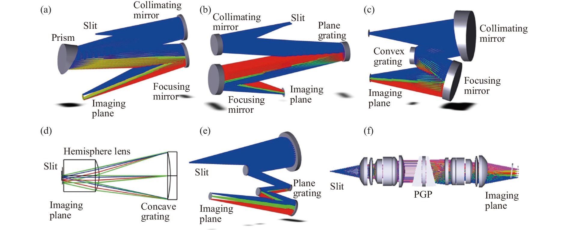
色散型成像光谱仪是目前所有成像光谱仪器中形式最为成熟的,也因为其高性能和高环境适应性成为了应用最为广泛的成像光谱仪类型。色散型成像光谱仪现已成为航空、航天、地面、工业、实验室研究等应用领域中的高精度定量探测的主要手段,发挥着举足轻重的作用。顾名思义,色散器件就是将入射的复合白光衍射为不同的单色光的核心光学元件,这种元件主要包括棱镜、光栅及其组合的分光器件。色散型成像光谱仪的基本组成包括狭缝、准直镜、色散分光器件、聚焦镜和探测器。
-
棱镜型成像光谱仪使用棱镜作为核心分光器件,其分光原理是组成棱镜的透射材料对不同波长具有不同的折射率,在棱镜的楔角α确定的情况下,一束光的出射偏向角θ与入射角i1的关系可以表示为[24]:
$$ \theta = {i_1} + {\arctan}\left( {n\sin \left( {\alpha - {\arctan}\left( {\frac{{\sin {i_1}}}{n}} \right)} \right)} \right) - \alpha $$ (4) 由公式(4)可知,当入射角i1不变时,偏向角θ即为折射率n的函数,因此偏向角θ也会随着波长的变化而变化,从而在通过棱镜折射后在空间上被分离色散开来。折射率一般随着波长的减小而增大,因此波长越短偏向角越大。
棱镜型成像光谱仪是各类色散型成像光谱仪中实施起来相对最容易的,结构也较为简单,其优点是可以在宽光谱下进行工作,工作带宽可以达到几百纳米甚至几微米;缺点是色散率低,导致仪器的光谱分辨率难以做到很高,且光谱分辨率在整个工作波段上并不均一分布,而是随着工作波长的增大而增大。图3(a)给出了一般棱镜型成像光谱仪的结构形式。
-
较为常见的光栅型成像光谱仪有使用平面反射式光栅的Czerny-Turner成像光谱仪,使用凸面和凹面光栅的同心类成像光谱仪(主要为Offner和Dyson成像光谱仪),以及使用棱镜和透射光栅共同组成核心分光器件的PG或PGP型成像光谱仪。
光栅基本方程表述为:
$$ \sin i \mp \sin\theta = mg\lambda $$ (5) 式中:i为入射角;θ为衍射角;m为光栅衍射级次;g为光栅刻线密度;λ为对应波长。光栅的色散率很高,因此光栅型成像光谱仪可以达到很高的光谱分辨率,但是由于光栅存在多级衍射效应,光栅型成像光谱仪的工作光谱不能过宽(级次滤光片可以一定程度上解决这一问题),另外精密光栅的制作极为复杂,光谱仪的装调也比棱镜型成像光谱仪的装调更为复杂。
-
Czerny-Turner光谱仪结构使用平面反射式光栅,其光谱分辨率可以做到很高。传统的Czerny-Turner光谱仪结构存在着一定的彗差和较大的像散,一般只在中心波长处具备良好的光学性能,因此常用于各类单色仪中,通过光栅转动实现不同的单色光输出。通过研究发现,对Czerny-Turner的准直镜和聚焦镜的形式进行改变[25-26],使用超环面镜或柱面镜来代替原来的球面镜形式,即可使其子午面和弧矢面上的曲率半径产生变化,同时延长光栅到聚焦镜的距离,就可以在较宽的工作波段上实现各像差(尤其是像散)的校正条件统一,从而对像差进行良好的抑制。这种改进型的Czerny-Turner成像光谱仪结构如图3(b)所示。
-
同心类成像光谱仪顾名思义其内部的所有光学元件的曲率半径中心位于同一点上,这种放置方法充分利用了Rowland圆的基本原理,使得系统可以非常充分地对各类像差进行抑制和消除,并可通过较高的数值孔径实现较大的光通量。同心类成像光谱仪主要有两种基本形式:一是Offner成像光谱仪结构[27];二是Dyson成像光谱仪结构[28],前者在1975年被首次提出,并被美国JPL实验室率先在工程上实现,成为目前航空航天中应用的各类先进高光谱成像仪的主要核心形式。Offner成像光谱仪主要由球面准直镜、凸面光栅和球面聚焦镜组成,其基本形式如图3(c)所示。在此基础之上又有很多种变形:包括在光路中增加弯月透镜、使用Littrow-Offner结构形式等不同的方法以进一步提高系统的数值孔径和压缩系统体积[29]。
Dyson成像光谱仪的产生时间要早于Offner成像光谱仪,其主要组成为半球透镜和凹面光栅,由于其满足Rowland圆的条件十分苛刻,导致狭缝和像面距离光学元件距离过近,因此在工程实施上具有极大的困难,故而较少得到应用,传统Dyson成像光谱仪结构如图3(d)所示。为了进一步发挥Dyson成像光谱仪的良好性能和应用,后来的很多研究人员都对Dyson成像光谱结构进行了一定程度的改进:目前最新的研究完成了Dyson系统的准同心条件分析,从而实现了改进型Dyson成像光谱仪的应用[30-32]。得益于Dyson结构的高通量、高成像质量的优点,Dyson成像光谱仪被应用于海洋水色成像、星载地物高光谱成像等领域。
此外,还有一种Schwarzschield成像光谱仪结构[33],它利用了Schwarzschield 光学系统(也称逆卡塞格林系统)作为准直镜和聚焦镜,并在中间使用平面光栅作为分光器件,其整体结构消除像差的原理与同心光谱仪类似,如图3(e)所示。
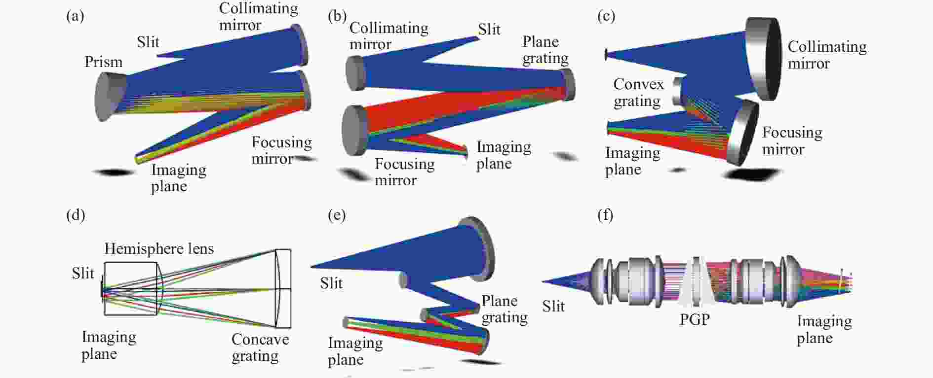
图 3 色散型成像光谱仪:(a) 棱镜成像光谱仪;(b) Czerny-Turner成像光谱仪;(c) Offner成像光谱仪;(d) Dyson成像光谱仪;(e) Schwarzschield 成像光谱仪;(f) PGP成像光谱仪
Figure 3. Dispersive imaging spectrometers: (a) Prism imaging spectrometer; (b) Czerny-Turner imaging spectrometer; (c) Offner imaging spectrometer; (d) Dyson imaging spectrometer; (e) Schwarzschield imaging spectrometer; (f) PGP imaging spectrometer
-
PGP型成像光谱仪是另一种相对比较常见的分光型成像光谱仪[34],如图3(f)所示,这种光谱仪的准直镜和聚焦镜均由透镜组构成;其核心器件是由贴合在一起的棱镜-透射光栅-棱镜(Prism-Grating-Prism)组成,这种组合色散元件的优点是很好地消除了一般透射光栅所带来的光谱谱线弯曲的问题[35],并且由于两端的棱镜使得光栅衍射分光产生的光路偏折得到了校正,使其光轴基本保持在同一条直线上,因此这种成像光谱仪在体积和形状上都较为规则,适于在各类小型观测平台上搭载,并且其数值孔径可以做的很高,适用于可见光-近红外等波段。
-
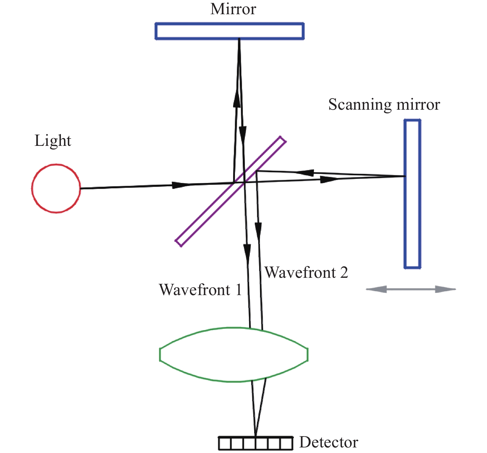
干涉型成像光谱仪(一般指傅里叶变换光谱仪)的基本原理是利用了具备光程差的波长干涉效应[36]。干涉型成像光谱仪的核心分光器件是分束器,将入射光束分成反射光和透射光两部分;传统傅里叶变换光谱仪(即迈克尔逊干涉仪)利用动镜产生不同的光程差,具有光程差的分束光线被后端聚焦镜组进行合束,并被探测器接收。通过对探测到的干涉图进行解析获取探测目标的特征光谱信息和成像信息,其光谱分辨率极高,干涉型成像光谱仪基本原理如图4所示。
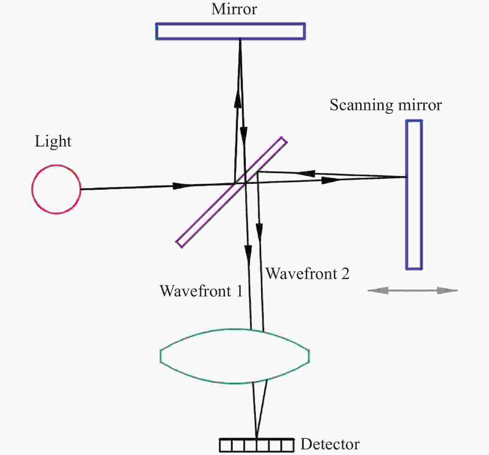
图 4 干涉型成像光谱仪原理示意图
Figure 4. Principle diagram of interferometric imaging spectrometers
干涉型成像光谱仪在获取数据立方体的过程中类似滤光型成像光谱仪,但是其光谱分辨率一般远高于滤光型成像光谱仪,且其获取探测目标全部光谱信息的速度要比色散型成像光谱仪快许多倍,因此具有很好的应用优势。但是干涉型成像光谱仪的系统精度和灵敏度也较高,因此对应用环境的要求较高,在具体的工程化实施中相比其他各类成像光谱仪难度最大。研究人员在传统迈克尔逊干涉成像光谱仪的基础上,研发了一些具备相似原理但又有所改进的成像光谱仪形式,比如:将上述的迈克尔逊干涉成像光谱仪内部的反射镜更改为内反射二面体,从而避免内部运动部件的使用,且对外界的干扰灵敏度更低;将反射镜进行一定角度的放置,从而产生更大的光程差,这种类型被称为Sagnac型成像光谱仪[37]。Mach-Zehnder (M-Z)型成像光谱仪[38-39]其分光器件采用两种不同材料的玻璃进行粘接,粘接界面处产生光的分束,分光器件出射面则为全反射,最终出射臂处产生不同光程差的光线,这种类型的成像光谱仪具备极高的环境适应性;另外,M-Z型干涉仪还可以通过电子调谐方式实现,因此非常适用于微型成像光谱仪的应用。此外,还有一些改进方法,比如:将反射镜替换为光栅,使用沃拉斯顿棱镜等偏振器件产生光程差等办法,以取代传统迈克尔逊干涉仪内部动镜,进一步提高仪器的环境适应性。
在星载、机载和地面的实际工程应用中,干涉型成像光谱仪因其高光谱分辨率和紧凑的体积等优点而逐渐得到广泛应用。
-
快照型成像光谱仪本质上是对以上各种成像光谱仪的一种进化改型,通过对分光器件或是孔径光阑的处理和改进使得成像光谱仪具备在一定范围内同时获得三维数据立方体的能力。目前主要的快照型成像光谱仪主要有以下几种。
-
这种类型的光谱仪是基于计算机生成的全息图色散元件(CGH: computer-generated hologram)设计而成[40]。在传统光谱仪前端物镜成像处不再放置狭缝,而是放置可通过较大光通量的光阑,利用全息色散元件(如:Kinoform光栅等)将混杂在一起的光谱信息和空间信息在探测器像面上的不同方向上进行投影,然后再利用CT重建算法获取观测目标的三维数据立方体(如图5(a)所示)。其优点是狭缝处为光阑,因此可以直接通过凝视来获得一个观测场景下的所有成像和光谱信息,而无需推扫或其他运动部件。计算层析成像光谱仪目前已经有了较为广泛的应用,在军事动态目标监测、生物医学临床图像结构获取、以及天文行星空间监测等领域都有建树。计算层析成像光谱仪的主要问题在于不容易获得较高的光谱分辨率,且在由低维投影数据计算高维原始图像过程中会产生一定的信息损失而影响重建图像的质量;另外,重建图像过程的实时性相对较差。
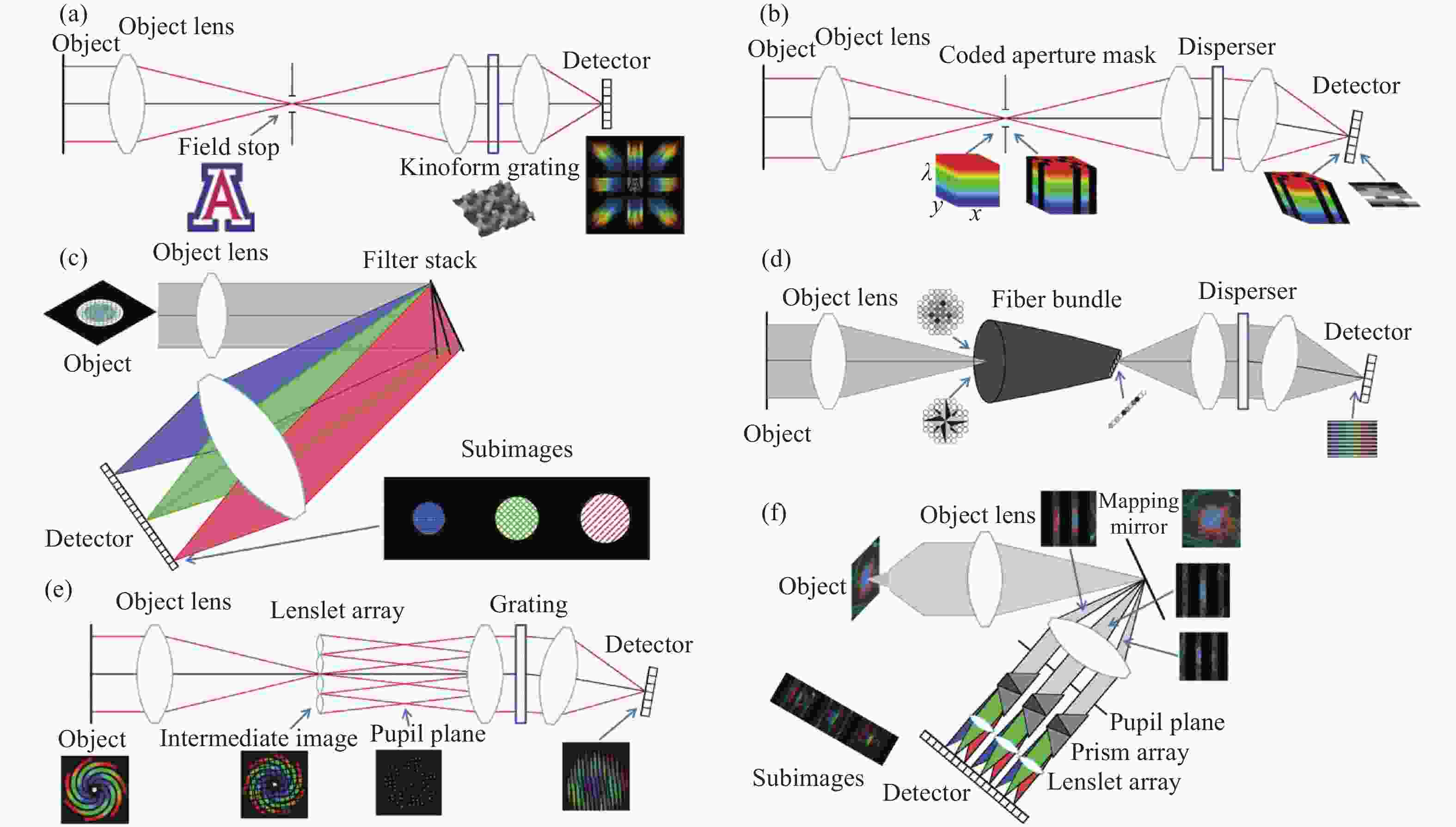
图 5 快照型成像光谱仪:(a) 计算层析成像光谱仪;(b) 编码孔径快照成像光谱仪;(c) 滤光片堆栈式解析成像光谱仪;(d) 光纤重组成像光谱仪;(e) 微透镜阵列场积分成像光谱仪;(f) 映射式成像光谱仪
Figure 5. Snapshot imaging spectrometers: (a) Computed tomographic imaging spectrometer (CTIS); (b) Coded aperture snapshot imaging spectrometer (CASSI); (c) Filter stack spectral decomposition (FSSD) imaging spectrometer; (d) Fiber-reformatting imaging spectrometer (FRIS); (e) Integral field spectroscopy with lenslet arrays (IFS-L) imaging spectrometer; (f) Image mapping imaging spectrometer (MSI)
-
这种成像光谱仪使用编码孔径板(通常为随机生成的二维二进制数字编码板)代替传统色散成像光谱仪中的狭缝,后端的色散成像系统对经过编码板编码后的图像进行接收和色散成像,并将观测场景的连续光谱图像投影在探测器上[41]。研究人员通过图像稀疏矩阵解析等方法对探测器接收信息进行解码获得最终所需的观测图像重建,原理如图5(b)所示。编码孔径成像光谱仪的核心器件为编码孔径器件,一般有移动式机械模板、液晶空间光调制器、数字微镜器件(DMD),其中DMD是目前应用较多的编码孔径器件,兼具数字电路灵活可控性与理想二值编码加载的优点。
编码孔径计算成像光谱仪是当今的成像光谱仪发展热门之一,其观测效率高,获取信息量大,具有很好的应用前景。但是这类光谱仪的工程化应用还需要更好的解决几个核心问题,包括:具备高精度和高环境适应性的二维孔径编码板的制造,具备高时间分辨率的实时解码算法的发展,光谱分辨率与解码算法之间的矛盾(具有高色散率的色散元件的应用会快速提升解码算法的复杂度)等。
-
滤光片堆栈成像光谱仪顾名思义就是使用一系列彼此存在一定夹角的窄带滤光片堆栈组代替单一的滤光片或滤光片轮,进而提高传统的多光谱相机时间分辨率,在同一观测时间内获取观测场景的多光谱成像信息[42],如图5(c)所示。这种形式的成像光谱仪可以代替传统的多光谱相机应用在各类测绘场景中。但是这种滤光片堆栈式的成像光谱仪同时获取的光谱通道数无法达到很多,且多个滤光片安装而成的堆栈对于后端的成像镜组要求较高,当滤光片较多时成像镜组和探测器像面均要求很大。
-
光纤重组成像光谱仪借助光纤束实现对前端物镜的成像信息接收和后端的整形图像输出[43]。光纤束将前端接收的信息通过光纤合束实现一维的信息排列,该信息被后端色散成像系统接收并最终成像在像面上,原理如图5(d)所示。此类成像光谱仪目前也开始得到一定的应用,其主要需解决的问题是减小光纤合束重组时信息和光通量的损失。
-
微透镜阵列场积分成像光谱仪使用微透镜阵列对前置物镜所成的中间像进行细分,再通过中间孔径后被后端色散成像光谱系统接收并最终成像在探测器像面上[44-45]。此类成像光谱仪的主要成像能力取决于微透镜阵列的性能参数,如图5(e)所示。微透镜阵列积分视场成像光谱仪最早在天文领域进行应用,可以通过一次曝光实现二维目标三维立方体数据采集,从而对天体进行观测,在国外的大部分大型望远镜上基本都配备有微透镜阵列积分视场成像光谱仪。
与微透镜阵列场积分成像光谱仪原理类似的另一种成像光谱仪的形式为细分成像切片镜场积分成像光谱仪,采用在一个反射镜上加工出不同楔角的长切片实现对图像的场重建。微透镜阵列场积分成像光谱仪的核心关键技术是微透镜阵列的制造,因此不能做的太大,且性能受限于微透镜阵列自身的性能;而细分成像切片镜的加工难度极大,目前仅有极少数国家具备同类元器件的加工技术,且价格十分昂贵。
-
映射式成像光谱仪通过中间的映射镜将图像切分为不同的条状场景[46],并且综合利用了棱镜阵列和微透镜阵列将不同切分场景进行色散成像,从而在探测器上获得一系列子图像,再通过算法将各子图像进行融合重建即可获得最终的高光谱图像立方体数据。此类成像光谱仪在元器件制作和整体集成上都有一定的难度,且光谱分辨率也不易做到很高。
快照型成像光谱仪所存在的共性问题主要在于其所获取的数据量是所有成像光谱仪中最大的,因此其所需解码和图像重建的数据量也是最大的,解码过程和处理算法比较复杂,在进行图像数据重建时耗时长,因此实时性较差。另外,快照型成像光谱仪很难做到极高的光谱分辨率,这是由于高光谱图像重建的难度与光谱分辨率的高低与光谱通道数成正比,很难实现兼顾。因此快照式成像光谱仪目前还没有完全进入成熟时期,需要进一步的开发和研究。
-
每一种成像光谱仪形式的发展都伴随着设计技术和制造技术的突破,并会针对其具体应用场景而进行进一步的改进和优化。无论是已有的成熟成像光谱仪形式,还是正在发展的新型成像光谱仪形式,都必然会随着科学技术的提升而诞生出新的活力,因此在宏观意义上讲各种形式并没有明显的优劣之分,只能说在特定的应用领域中,某一类型的成像光谱仪其工作方式和性能更为突出。当然,尽管不同类型的成像光谱仪在性能上有所侧重,但是所有的成像光谱仪在通用指标上的未来发展方向都是向着高光谱分辨率、高空间分辨率、高光通量和高时间分辨率发展的。
滤光型成像光谱仪本质上即为高光谱相机,也是最早在工程中应用的成像光谱仪类型之一,目前技术相对最为成熟,尤其在具备超高空间分辨率的多光谱详查和测绘相机载荷中应用最为广泛。在所有的成像光谱仪形式中,滤光型成像光谱仪一般而言是体积最小和最为紧凑的,覆盖场景能力(视场角)也是最大的,因此在有高机动性要求的小型平台(如:无人机平台等)上搭载可以发挥巨大的作用。在未来,无论是星载平台还是机载平台,滤光型成像光谱仪仍然是重要的仪器应用类型之一。另外,随着制造技术的不断发展,干涉式滤光片的线宽将可进一步减小达到与色散型成像光谱仪的光谱分辨能力同等的程度,而随着各种光学可调谐器件的进一步发展,其光谱分辨能力也将获得进一步提高,达到甚至超过干涉型成像光谱仪的光谱分辨率,因此滤光型成像光谱仪也将随着各种先进技术的发展而进一步焕发青春。
色散型成像光谱仪一直是成像光谱仪的主要代表形式和应用形式,这种类型的成像光谱仪从单色仪发展而来,且环境适应性很高,因此也是目前各类星载、机载、船载、地基高光谱成像仪的主流结构形式。国内外的多个星上高精度定量观测载荷均采用了色散型成像光谱仪的形式,可以说色散型成像光谱仪的发展已经进入了成熟阶段。随着制造技术和设计技术的进一步发展,变线间距光栅、特殊曲面光栅、特殊曲面棱镜等特殊色散元件逐渐开始得到应用,这势必将为色散型成像光谱仪的性能进一步发展提供技术基础。我国特种光栅的制造生产能力还相对薄弱,一定程度上影响了具备自主知识产权的色散型成像光谱仪的研制和发展;另外,我国的高光谱成像仪商业化发展也比较滞后,而色散型成像光谱仪在各类科研工作中是存在较大缺口的,使用产品主要以国外厂商的设备为主,需要进行产业化的关键技术攻关与发展。
干涉型成像光谱仪在各类高精度探测和校准仪器中得到了广泛应用。除了应用于星上和飞行平台进行大气、风场等环境参数的高精度监测外,另一大重要应用就是地面检测校准仪器,如:Zygo和4D干涉仪等。中国科学院西安光学精密机械研究所、中国科学院安徽光学精密机械研究所和中国科学院空天信息创新研究院在此领域具有很好的研究基础,其自主开发的仪器除了在卫星平台上得到应用外,也开始在地面上应用于实验室检测校准。
快照式成像光谱仪是近年来开始得到全面研究的新兴研究领域,这种成像光谱仪在传统成像光谱仪的基础上综合了数学、物理、微纳器件、图像算法甚至深度学习等多学科知识,力图在最大程度上快速、完整地获得整个探测场景的三维立方数据体。虽然目前这一领域还存在着一些技术瓶颈,但是在未来,快照式成像光谱仪必将在军事应用、环境监测、目标识别领域得到应用。
成像光谱仪发展的另一个重要方向是微小型化的应用,即将分光能力和器件最大程度地集成化和小型化。目前基于MEMS的可调谐滤光型成像光谱仪、傅里叶变换干涉型成像光谱仪和光栅式的色散型成像光谱仪已经基本实现了商业化,部分仪器甚至已经实现了消费级应用。随着相关技术的发展,各类微小型成像光谱仪的性能还可以得到进一步提升。而在未来,随着快照型计算式成像光谱技术的进一步发展,有望将已达到极小体积(百微米级别)的核心器件和设备缩小到10 μm级别。
成像光谱技术是一门融合了传统光学和新兴技术的特殊学科,且将随着人类需求的提升和技术的发展而不断焕发出新的生命力。我国的成像光谱仪虽然起步较晚,但是在仪器核心设计、研制、集成和应用方面都得到了很好的发展,很多领域都处于国际领先地位。该领域从业者需进一步加强该领域的基础性前沿研究,强化我国在该领域的研究基础;同时提高核心元器件的自主设计制造能力,提升发展应用水平,打破国外的工业化垄断。
Development and application of imaging spectrometer (Invited)
-
摘要: 成像光谱仪是一种有效的定量探测工具,得益于其可通过高空间分辨率和高光谱分辨率同时获取探测目标的三维立方体数据的能力,使得成像光谱仪在测绘遥感、目标识别、环境监测与评估、临床影像诊断、过程监控等领域应用极为广泛。文中根据分光原理和分光元件的不同,将成像光谱仪分为四类:滤光型、色散型、干涉型和快照型,在分类的基础上对各种成像光谱仪的主要代表形式和应用进行了回顾和介绍,并对成像光谱仪未来可能存在的发展和应用方向进行了展望。Abstract: Imaging spectrometers, which collect data over three dimension-two spatial and one spectral so that a complete dataset is typically referred to as a datacube, is one of the most powerful and widely used characterization tools in accurate mapping of wide areas remote sensing, object identification and recognition, environment assessment and management, clinical diagnosis imaging and process monitoring and control. According to the different spectral principle and component, imaging spectrometers were divided into four classes as filtering imaging spectrometers, dispersive imaging spectrometers, interferometric imaging spectrometers and snapshot imaging spectrometers. A comprehensive review of main representative forms and applications of various imaging spectrometers on the basis of classification was provided, and the possible development and application of imaging spectrometers in the future were prospected.
-
Key words:
- imaging spectrometer /
- filter /
- dispersion /
- interference /
- snapshot
-
图 1 不同成像光谱仪在单次观测积分时间下获取三维体立方数据过程示意图
Figure 1. Portions of the datacube collected during a single observing integration period for different imaging spectrometers
图 2 滤光型成像光谱仪主要分光原理:(a) 干涉式滤光片;(b) 声光可调谐滤光器;(c) Lyot-Ohman液晶双折射可调谐滤光器;(d) 法布里-珀罗腔;(e) 阵列式滤光器; (f) 线性渐变式滤光片
Figure 2. Main principle of filter imaging spectrometers: (a) Interference filter;(b) Acousto-optic tunable filter;(c) Lyot-Ohman liquid crystal birefingent tunable filter;(d) Fabry-Perot etalon;(e) Array filter; (F) Linear variable filter
图 3 色散型成像光谱仪:(a) 棱镜成像光谱仪;(b) Czerny-Turner成像光谱仪;(c) Offner成像光谱仪;(d) Dyson成像光谱仪;(e) Schwarzschield 成像光谱仪;(f) PGP成像光谱仪
Figure 3. Dispersive imaging spectrometers: (a) Prism imaging spectrometer; (b) Czerny-Turner imaging spectrometer; (c) Offner imaging spectrometer; (d) Dyson imaging spectrometer; (e) Schwarzschield imaging spectrometer; (f) PGP imaging spectrometer
图 4 干涉型成像光谱仪原理示意图
Figure 4. Principle diagram of interferometric imaging spectrometers
图 5 快照型成像光谱仪:(a) 计算层析成像光谱仪;(b) 编码孔径快照成像光谱仪;(c) 滤光片堆栈式解析成像光谱仪;(d) 光纤重组成像光谱仪;(e) 微透镜阵列场积分成像光谱仪;(f) 映射式成像光谱仪
Figure 5. Snapshot imaging spectrometers: (a) Computed tomographic imaging spectrometer (CTIS); (b) Coded aperture snapshot imaging spectrometer (CASSI); (c) Filter stack spectral decomposition (FSSD) imaging spectrometer; (d) Fiber-reformatting imaging spectrometer (FRIS); (e) Integral field spectroscopy with lenslet arrays (IFS-L) imaging spectrometer; (f) Image mapping imaging spectrometer (MSI)
-
[1] Mouroulis P, Green R O. Review of high fidelity imaging spectrometer design for remote sensing [J]. Optical Engineering, 2018, 57(4): 040901. [2] Lumb D H, Bautz M W, Burrows D N, et al. Recent developments for the AXAF CCD imaging spectrometer [C]//EUV, X-Ray, and Gamma-Ray Instrumentation for Astronomy IV, International Society for Optics and Photonics, 2006: 265-271. [3] Kudenov M W, Dereniak E L. Compact real-time birefringent imaging spectrometer [J]. Optics Express, 2012, 20(16): 17973-17986. doi: 10.1364/OE.20.017973 [4] Wang R S, Yang S M, Yan B. A review of mineral spectral identification methods and models with imaging spectrometer [J]. Remote Sensing for Land & Resources, 2007, 19(1): 1-9. [5] Gons H J, Rijkeboer M, Ruddick K G. A chlorophyll-retrieval algorithm for satellite imagery (medium resolution imaging spectrometer) of inland and coastal waters [J]. Journal of Plankton Research, 2002, 24(9): 947-951. doi: 10.1093/plankt/24.9.947 [6] Schlerf M, Atzberger C, Hill J. Remote sensing of forest biophysical variables using HyMap imaging spectrometer data [J]. Remote Sensing of Environment, 2005, 95(2): 177-194. doi: 10.1016/j.rse.2004.12.016 [7] Lerner J M. Imaging spectrometer fundamentals for researchers in the biosciences—A tutorial [J]. Cytometry Part A: the Journal of the International Society for Analytical Cytology, 2006, 69(8): 712-734. [8] Warren M A, Simis S G H, Martinez-Vicente V, et al. Assessment of atmospheric correction algorithms for the Sentinel-2A multispectral imager over coastal and inland waters [J]. Remote Sensing of Environment, 2019, 225: 267-289. doi: 10.1016/j.rse.2019.03.018 [9] Beutler H G. The theory of the concave grating [J]. J Opt Soc Am, 1945, 35(5): 311-350. [10] Werner W. The geometric optical aberration theory of diffraction gratings [J]. Applied Optics, 1967, 6(10): 1691-1699. doi: 10.1364/AO.6.001691 [11] Mathew S K, Bayanna A R, Tiwary A R, et al. First observations from the Multi-Application Solar Telescope (MAST) narrow-band imager [J]. Solar Physics, 2017, 292(8): 1-25. [12] Xu Z, Zhao H, Jia G, et al. Optical schemes of super-angular AOTF-based imagers and system response analysis [J]. Optics Communications, 2021, 498(8): 127204. [13] Yamashita T, Kinoshita H, Sakaguchi T, et al. Objective tumor distinction in 5-aminolevulinic acid-based endoscopic photodynamic diagnosis, using a spectrometer with a liquid crystal tunable filter[J]. Annals of Translational Medicine, 2020, 8(5): 178. [14] Antila J, Miranto A, Mkynen J, et al. MEMS and piezo actuator-based Fabry-Perot interferometer technologies and applications at VTT [C]//Proceedings of SPIE - The International Society for Optical Engineering, 2010, 7680: 76800U. [15] Prieto-Blanco X, Montero-Orille C, Couce B, et al. Optical Configurations for Imaging Spectrometers [M]//Graña M, Duro R J. Computational Intelligence for Remote Sensing. Heidelberg, Berlin: Springer, 2008: 1-25. [16] Jerman J H , Clift D J , Mallinson S R. A miniature Fabry-Perot interferometer with a corrugated silicon diaphragm support [C]//IEEE Solid-state Sensor & Actuator Workshop, IEEE, 1991, 29(2): 151-158. [17] Correia J H, Graaf G, Kong S H, et al. Single-chip CMOS optical microspectrometer[J]. Sensors and Actuators A: Physical, 2000, 82(1-3): 191-197. [18] Wang S W, Xia C, Chen X, et al. Concept of a high-resolution miniature spectrometer using an integrated filter array [J]. Optics Letters, 2007, 32(6): 632-634. doi: 10.1364/OL.32.000632 [19] Bryan K M, Jia Z, Pervez N K, et al. Inexpensive photonic crystal spectrometer for colorimetric sensing applications [J]. Optics Express, 2013, 21(4): 4411-4423. doi: 10.1364/OE.21.004411 [20] Hang Q, Ung B, Syed I, et al. Photonic bandgap fiber bundle spectrometer [J]. Applied Optics, 2010, 49(25): 4791-4800. doi: 10.1364/AO.49.004791 [21] Tittl A, Leitis A, Liu M, et al. Imaging-based molecular barcoding with pixelated dielectric metasurfaces [J]. Science, 2018, 360(6393): 1105-1109. doi: 10.1126/science.aas9768 [22] Cheriton R, Densmore A, Dezfouli M K, et al. Exoplanetary atmosphere spectroscopy using silicon waveguide ring resonators [C]//2019 Photonics North (PN), IEEE, 2019: 1-1. [23] Mika. Linear-wedge spectrometer [C]//Imaging Spectroscopy of the Terrestrial Environment, International Society for Optics and Photonics, 1990, 1298: 127-131. [24] Tang Z, Gross H. Improved correction by freeform surfaces in prism spectrometer concepts [J]. Applied Optics, 2021, 60(2): 333-341. doi: 10.1364/AO.412103 [25] Dong J, Chen H, Zhang Y, et al. Miniature anastigmatic spectrometer design with a concave toroidal mirror [J]. Applied Optics, 2016, 55(7): 1537-1543. doi: 10.1364/AO.55.001537 [26] Zhao Yiyi, Yang Jianfeng, Xue Bin, et al. Optical system design of broadband astigmatism-free Czerny-Turner spectrometer [J]. Infrared and Laser Engineering, 2014, 43(4): 1182-1187. (in Chinese) [27] Offner A. New concepts in projection mask aligners [J]. Optical Engineering, 1975, 14(2): 142130. [28] Dyson J. Unit magnification optical system without Seidel aberrations [J]. Josa, 1959, 49(7): 713-716. doi: 10.1364/JOSA.49.000713 [29] Wu S, Huang C, Yu L, et al. Analytical design of an advanced Littrow–Offner spectrometer simultaneously off-axis in meridian and sagittal planes for ultraviolet imaging with high performance [J]. Applied Optics, 2021, 60(32): 10094-10100. doi: 10.1364/AO.441758 [30] Yu L. Upgrade of a UV-VIS-NIR imaging spectrometer for the coastal ocean observation: concept, design, fabrication, and test of prototype [J]. Optics Express, 2017, 25(13): 15526-15538. doi: 10.1364/OE.25.015526 [31] Lei Y, Sujuan C, Jiexiang C, et al. Optical system of imaging spectrometer in short-wave infrared with high NA for precision agriculture observation [J]. Infrared and Laser Engineering, 2018, 47(12): 1218007. (in Chinese) doi: 10.3788/IRLA201847.1218007 [32] Wu S, Huang C, Yu L, et al. Optical design and evaluation of an advanced scanning Dyson imaging spectrometer for ocean color [J]. Optics Express, 2021, 29(22): 36616-36633. doi: 10.1364/OE.440840 [33] Mouriz M D, Lago E L, Prieto-Blanco X, et al. Schwarzschild spectrometer [J]. Applied Optics, 2011, 50(16): 2418-2424. doi: 10.1364/AO.50.002418 [34] Xue Q, Qi M, Li Z, et al. Fluorescence hyperspectral imaging system for analysis and visualization of oil sample composition and thickness [J]. Applied Optics, 2021, 60(27): 8349-8359. doi: 10.1364/AO.432851 [35] Zhikun Wu, Entao Shi, Yongmei Wang. Design of PGP imaging spectrometer with eliminating spectral line bending [J]. Infrared and Laser Engineering, 2021, 50(6): 20200433. (in Chinese) [36] Pisani M, Zucco M. Compact imaging spectrometer combining Fourier transform spectroscopy with a Fabry-Perot interferometer [J]. Optics Express, 2009, 17(10): 8319-8331. doi: 10.1364/OE.17.008319 [37] Rafert J B, Sellar R G, Blatt J H. Monolithic Fourier-transform imaging spectrometer [J]. Applied Optics, 1995, 34(31): 7228-7230. doi: 10.1364/AO.34.007228 [38] Xiao Xiangguo, Zhang Shuanmin, Chen Xiuping. Image plane intersection interference imaging spectrometer based on Mach-Zehnder [J]. Infrared and Laser Engineering, 2016, 45(5): 0524001. (in Chinese) [39] Yang M, Li M, He J J. Static FT imaging spectrometer based on a modified waveguide MZI array [J]. Optics Letters, 2017, 42(14): 2675-2678. doi: 10.1364/OL.42.002675 [40] Hagen N, Dereniak E L. New grating designs for a CTIS imaging spectrometer [C]//Algorithms and Technologies for Multispectral, Hyperspectral, and Ultraspectral Imagery XIII, International Society for Optics and Photonics, 2007, 6565: 65650N. [41] Pei L L, Lv Qunbo, Wang Jianwei, et al. Optical system design of the coded aperture imaging spectrometer [J]. Acta Physica Sinica, 2014, 63(21): 210702. (in Chinese) [42] Shirokoff E, Barry P S, Bradford C M, et al. Design and performance of superspec: An on-chip, kid-based, mm-wavelength spectrometer [J]. Journal of Low Temperature Physics, 2014, 176(5): 657-662. [43] Uto K, Seki H, Saito G, et al. Development of a low-cost hyperspectral whiskbroom imager using an optical fiber bundle, a swing mirror, and compact spectrometers [J]. IEEE Journal of Selected Topics in Applied Earth Observations and Remote Sensing, 2016, 9(9): 3909-3925. doi: 10.1109/JSTARS.2016.2592987 [44] Liu Jianan, Cui Jichen, Yin Lu, et al. Analysis and design of pre-imaging system of integral field imaging spectrometer based on lenslet array [J]. Spectroscopy and Spectral Analysis, 2018, 38(10): 3269-3272. (in Chinese) [45] Liu J, Chen J, Liu J, et al. Optical design of a prism-grating-based lenslet array integral field spectrometer [J]. Optics Express, 2018, 26(15): 19456-19469. doi: 10.1364/OE.26.019456 [46] Yuan Y, Ding X M, Su L J, et al. Modeling and analysis for the image mapping spectrometer [J]. Chinese Physics B, 2017, 26(4): 040701. doi: 10.1088/1674-1056/26/4/040701 -

 点击查看大图
计量
- 文章访问数: 1075
- HTML全文浏览量: 252
- PDF下载量: 337
- 被引次数: 0


