


-
弹性是物质材料的基本物理性质,在生物医学及材料等研究中发挥着不可替代的作用,尤其在疾病诊断中,人体组织的弹性信息起到至关重要的作用。生物组织的不同分子及其在人体组织中的组织形式决定了它的弹性和硬度[1-2]。在人体组织中,不同形式的解剖结构弹性会有差异。一般情况下,正常组织之间的弹性有微小的差别,比如,在正常的乳房组织中,乳房组织的硬度要小于纤维组织的硬度,而又略大于脂肪组织。然而,对于部分特别的正常组织来说,其与变异组织的弹性差异比较明显,如肝转移癌、乳腺硬结癌等恶性病变通常以硬结节[3]的形式出现。乳腺硬结癌是最常见的乳腺癌,由于基体密度增加,呈现出致密的块状物。此外,其他一些病变也会改变组织的弹性系数,如肝纤维化、胶原沉积等疾病。肝活检是判断病人的肝脏组织是否纤维化的重要手段[4],由于肝活检是一种侵入性的检测方法,且肝纤维化是随机分布的,这不仅降低了测量结果的准确性,而且可重复性也很差,另外,肝活检的费用很高,普通病人很难承担。一般的医学检测方式,如X射线成像[5]、计算机断层扫描(CT)[6]、磁共振(MRI)[7]以及超声成像(USI)[8],无法直接提供组织的基本弹性信息。因此,研究一种能够无创、无标记、不接触、准确测量生物组织弹性模量的技术[9],预期可在未来的癌症、活体眼组织的表征和其他医学疾病的临床诊断中发挥重要作用。
目前,弹性成像和光声成像是获取生物组织弹性信息的常用方法。弹性成像技术获得的弹性信息的信噪比及对比度较低,无法提供可靠的病变边界信息[10]。相比之下,最新发展起来的布里渊光谱散射技术是一种光声成像技术,不仅分辨率高、无标记,而且是三维成像、不接触[11-12],因此可应用于临床诊断[13]、环境感知[14]和材料科学[15]等领域,受到了国内外众多专家学者的关注。随着激光技术的进一步发展,布里渊光谱学在材料的粘弹性和热力学性质研究中得到了广泛的应用[16]。2007年,Giuliano 和 Seoks首次将布里渊技术从点采样光谱扩展到成像模式[17],监测了聚合物交联过程中弹性模量的快速动态变化;2018年,Dana等人利用532 nm单频激光作为光源,使用Sandk级联的FP干涉仪和PMT测量了压缩后的牛骨弹性特性, 发现缺陷骨具有小的弹性模量[18]。光声技术多数是基于串联多通道的Fabry-Perot干涉仪方法进行布里渊散射频移测量,光谱分辨率可达0.5 GHz,但此方法系统组成庞大又复杂,实验过程以及后续的数据处理非常耗时,而且系统的造价昂贵,很难进行推广应用。
文中提出了一种基于虚像相位阵列的布里渊光谱的材料弹性模量测量方法,能够快速获得生物组织和材料的布里渊光谱频移,从而得到其弹性模量。由于虚像相位阵列一次分光谱的特性,所以这种测量方法不仅采集速度快,而且光谱分辨率高,可达0.25 GHz。单点光谱实验中,测得了聚乙烯、二氧化硅玻璃和PMMA样品的布里渊光谱频移,分别为16 GHz、33.8 GHz和15.4 GHz,求得样品材料的纵向弹性模量M分别为7.23 GPa、83.4 GPa和8.92 GPa。
-
共焦成像是一种采用点对点照明及空间针孔调制,从而消减样品在非焦平面散射光的光学成像方法,此方法具有很高的光谱分辨率[19]。
布里渊散射光谱是通过散射光的光谱偏移来研究样品中入射光和声学声子之间的相互作用,从而提供关于材料的声学,热力学和粘弹性特性的信息。在单一材料中,声学声速和布里渊光谱频移的关系为:
式中:
$\Delta \nu $ 为布里渊光谱的频移量;$\lambda $ 为入射波长;$V$ 为声学声速;${{n}}$ 为折射率;$\theta $ 为散射角。若是后向散射模式,则
$\theta $ =180°,由公式(1)可得:由参考文献[8]可知,单一材料的纵向弹性模量M为:
式中:
$\rho $ 为材料的密度。因此,通过公式(2)~(3),布里渊光谱在后向散射的情况下,通过测量材料的布里渊频移
$\Delta \nu $ ,并结合该材料的密度$\rho $ ,就可得到该材料的纵向弹性模量M。 -
虚像相位阵列(Virtual Image Phase Array, VIPA)实质上是一个表面粗糙度小于0.01的平行平板,它的基本原理如图1所示。入射面和出射面都被镀上高反膜,一般分别为100%和95%。入射光以小角度进入玻璃板并聚焦到出射面上,其中一小部分会穿过出射面,通过光束腰后发散,大部分的光被反射到入射面后会再次反射到出射面,然后再有一小部分光从出射面射出。如此反复,直到所有光泄露出VIPA。所有的出射光之间会产生相互干涉,被分成无数间隔固定的光束,而每一个光束都可以假设成是由一个虚像产生的。一般情况下,VIPA的光谱分辨率为[20]:

Figure 1. Basic principle of VIPA
式中:
${\theta _1}$ 为VIPA的倾角;$d$ 为VIPA 的厚度;${\lambda _0}$ 为激光中心波长;${{n}}$ 为空气折射率;${{{R}}_1}$ 、${R_2}$ 分别为VIPA前后表面膜的反射率。出射光束中的光由于波长不同,折射出的光角度各不相同,形成色散效应[21]。根据VIPA的色散效应,可以获得不同入射光的光谱特征。此外,由于VIPA是标准具,受到自由光谱范围(Free Spectral Range, FSR)的限制,会出现级次重叠现象,如图2所示,VIPA标准具无法区分550.00 nm与550.06 nm波长的光。因此,VIPA常与其他角度色散光学元件级联使用,不仅提高了采集速率和分辨率,而且能够改善级次重叠的现象,从而获得二维光谱图像。该系统中使用的VIPA是LightMachinery Inc.的OP-6721-2000-2Rev.F VIPA500~600 nm, 厚度为2 mm ,光谱分辨率为1 pm,同步测量范围为4 nm。

Figure 2. Orders overlap phenomenon of the VIPA
-
图3为共焦布里渊光谱成像系统,由光谱激发、白光照明、光谱采集、三维空间运动四个模块组成。首先,使用白光照明模块和三维空间运动实现被测样品的定焦与目标区域的确定;然后,利用光谱激发模块产生布里渊散射光,并收集后向散射光进入光谱采集模块,利用VIPA光谱仪采集被测目标的布里渊光谱信息,每个模块的具体组成和作用见3.1~3.4小节。

Figure 3. Confocal Brillouin spectral imaging system
-
光谱激发模块如图3中红色实线部分所示,它的作用是激发布里渊散射光。包含激光、会聚物镜、准直器扩散器、分束棱镜、显微物镜和共焦针孔等。激光器使用的是Verdi G-2W 532 nm单纵模、连续、大功率、光抽运半导体激光器,由相干公司生产。激光输出通过光束扩散器扩展光束,微元区域汇聚到焦平面上,实现“点光源”。焦平面处的微元物质和激光激发出的光子相互作用,从而激发出布里渊散射光,显微物镜将收集的背向散射光汇聚进入物镜A、直径为100 μm的共焦针孔及物镜B中,为了将针孔置于物镜A的物侧焦点处,实现“点检测”,需要反复调整针孔的位置。
-
图3中的白光照明模块设计用于确定被测样品的固定焦点和靶区,主要组件有CCD、分光棱镜、显微物镜和白光光源。CCD获得样品的靶区和样品的轴向聚焦,显微物镜进行显微成像。显微物镜使用的是Olympus生产的PLAN系列、数值孔径是0.25的10倍显微镜,其作用是白光聚焦成像。在进行样品光谱测量之前,使用白光照明模块。
-
图3所示的三维空间运动模块用于样品观测、靶区选择和布里渊光谱成像。平台分为横向(垂直于光轴) XY平面及轴向(光轴)。其中横向XY平面运动平台有两种调节方式,分别为粗调、精调,粗调二维平台使用的是BioPrecision2系列电动平移平台,分辨率为50 nm,行程为100 mm×100 mm。精控部分采使用P-542.2CD二维运动平台,行程为200 μm×200 μm,闭环分辨率与线性度为2 nm及0.03%,重复性小于5 nm。轴向运动部件使用的是SGSP80-20ZF型升降平台,用于样品定位过程中的轴向位置的调节。
-
布里渊弹性散射的频移大致在1~50 GHz范围内,所以光谱仪的分辨率必须满足R> 106的要求。现在已有的光谱检测方法有多通道扫描FP干涉仪法[22]、光栅单色仪法[23]等。然而,上述扫描方法不仅分辨率低而且采集速度慢,缓慢的采集时间将布里渊光谱限制为点采样或静态测量。针对上述问题,该系统使用基于虚像相位阵列(Virtual Image Phase Array,VIPA)的光谱仪作为布里渊光谱采集模块。图4为VIPA光谱仪的示意图,红色实线部分为激光和背向散色光抑制模块,红色虚线部分为二维光谱成像模块。

Figure 4. Schematic diagram of VIPA spectrometer
-
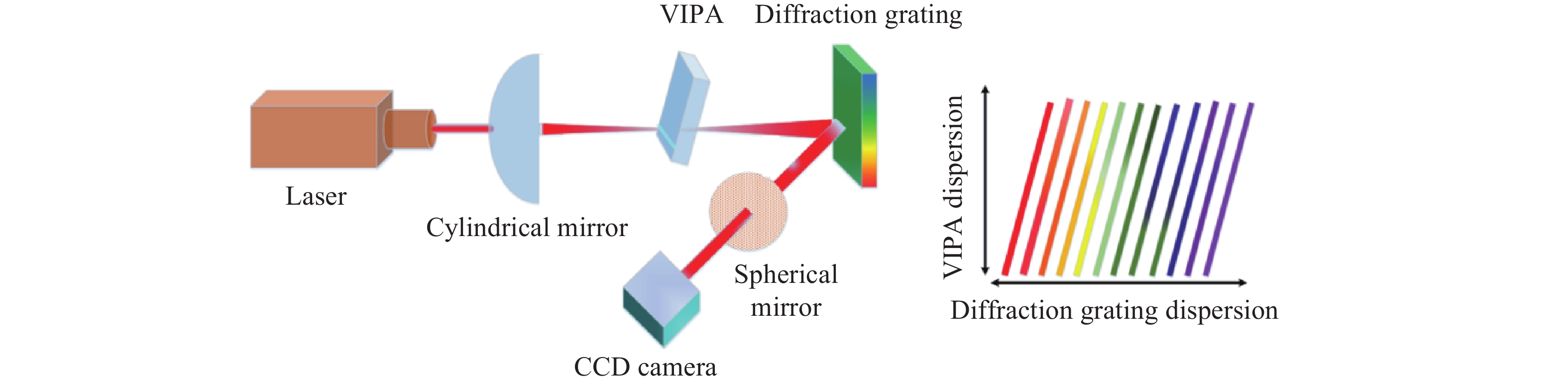
为了提高布里渊光谱的采集速度和光谱分辨率,该系统将VIPA与衍射光栅级联。图5为VIPA和衍射光栅联合的二维光谱原理图。垂直方向上,VIPA提供高分辨率;水平方向上,衍射光栅将重叠的标准具级次分开,以扩展整个高分辨率光谱。所有波长可以同时采集,大大缩短了光谱的采集时间。此外,将VIPA与衍射光栅结合,输出光束会根据不同的波长在二维平面展开,从而使得波长和成像面空间中的坐标一一对应,从而测量出物体的二维光谱图像信息。
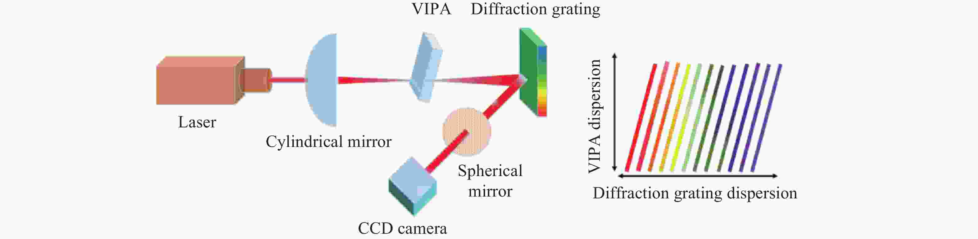
Figure 5. Two-dimensional spectral principle of VIPA combined with diffraction grating
-
布里渊散射在自发状态下是相对较弱的过程,并且光频移很小,大约为10 GHz。在大多数情况下,由瑞利散射或光学反射引起的弹性散射光比布里渊信号强几个数量级,因此弹性散射信号很容易淹没布里渊信号。为了改善这一情况,该方案采用超窄线宽滤波器来抑制激光在光谱仪内部的反射,其滤波原理与F-P标准具相似。F-P标准具透射率最大的条件为:
图6为超窄线宽滤波器原理图,根据不同波长
$\lambda $ ,通过调节可移动平板来改变间距${{d}}$ ,即可达到滤波的目的。
Figure 6. Schematic diagram of ultra-narrow line-width filter
-
为了检验该装置的光谱检测特性及成像功能,进行了单个点的光谱探测和二维光谱成像两个实验。
-
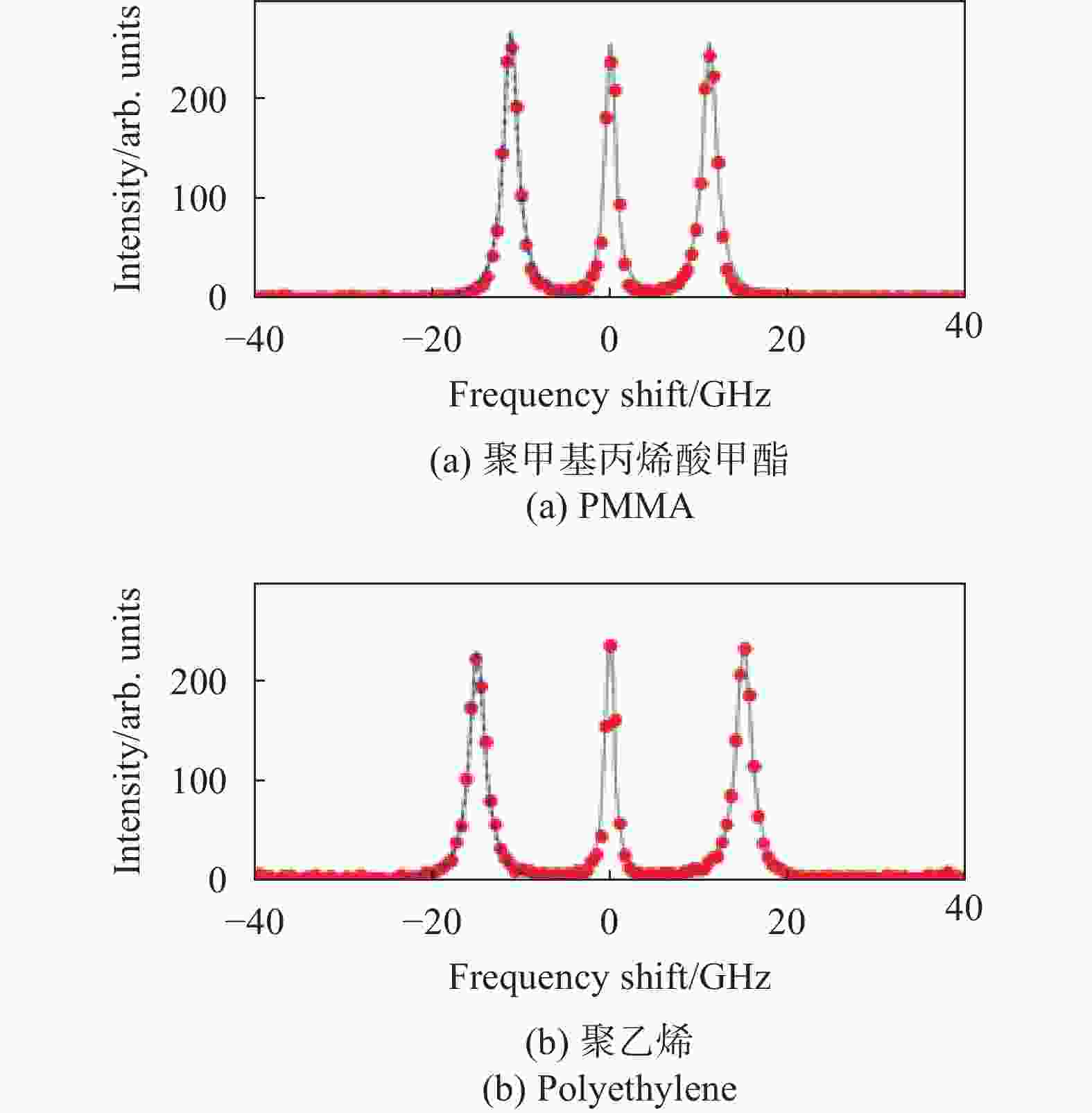
对聚甲基丙烯酸甲酯(PMMA)、聚乙烯和二氧化硅玻璃样品分别进行了单点的布里渊光谱测试。图7分别为PMMA、聚乙烯和二氧化硅玻璃样品的典型光谱仪输出图。

Figure 7. Typical spectrometer output of the sample
使用洛伦兹峰值拟合方法,获得了PMMA、聚乙烯和二氧化硅玻璃的布里渊光谱频移,分别为15.4 GHz、16 GHz和33.8 GHz,如图8所示,然后根据公式(2)及公式(3)计算得出上述各材料的纵向弹性模量M,如表1所示,实验结果与参考文献[12]测试结果一致。
Sample Density/
g·cm−3Refractive
indexFrequency
shift/GHzElastic
modulus/GPaPMMA 1.18 1.49 15.4 8.92 Polyethylene 0.19 1.51 16 7.23 Silica glass 2.23 1.47 33.8 83.4 Table 1. Typical parameters of PMMA, polyethylene and silica glass[12]

Figure 8. Brillouin spectrum of PMMA, polyethylene and silica glass
该系统布里渊光谱的分辨率计算式为[24]:
式中:
$\Delta {{f}}$ 为光谱分辨率;$c$ 为光速(3×10⁸ m/s);$N$ 为通道数(4096);$\Delta \lambda $ 是同步测量范围(4 nm);${\lambda _0}$ 为激光中心波长(532 nm)。代入测量参数,可以得到该实验装置的光谱分辨率为0.25 GHz。 -
由于容易获得PMMA的布里渊信号,因此在该实验中选择了PMMA来验证成像性能。图9是PMMA的二维布里渊光谱图像。图9(a)是在没有激光和反向散射光抑制模块的情况下测得的PMMA的布里渊光谱图像。图9(b)是添加了激光和反向散射光抑制模块后测得的PMMA的布里渊光谱图像,图中两个白色亮点即为PMMA的布里渊光谱图像。通过实验可知,加入激光和背向散色光抑制模块,可以更清楚更有效地获得物质的布里渊光谱图像信息。

Figure 9. Brillouin spectral image of PMMA
-
布里渊光谱技术具有高分辨率及非接触式等优点,已成为材料弹性研究的重要方法。利用该实验研制的布里渊光谱测量装置,测得了聚乙烯、二氧化硅玻璃和PMMA样品的布里渊光谱频移,分别为16 GHz、33.8 GHz和15.4 GHz,求得样品材料的纵向弹性模量M分别为7.23 GPa、83.4 GPa和8.92 GPa。此外,还测得了PMMA的布里渊光谱图像。该装置还可应用于组织损伤检测、材料特性表征等领域,对生物医学、材料科学、仿生学等方面的发展具有非常重要的作用。
Elastic modulus measurement based on virtual image phase array confocal Brillouin spectroscopy technology
doi: 10.3788/IRLA20200265
- Received Date: 2020-03-10
- Rev Recd Date: 2020-04-25
- Available Online: 2021-05-12
- Publish Date: 2021-04-30
-
Key words:
- elastic modulus /
- virtual image phase array /
- confocal Brillouin /
- spectral frequency shift
Abstract: The elastic modulus of biological tissues and materials is the key factor to determine their mechanical properties, which is very important for biomedicine and material monitoring. A method for measuring elastic modulus based on virtual image phase array was proposed. Combining virtual image phase array with confocal microscopy system, the Brillouin spectrum frequency shift of biological tissues and materials could be obtained quickly, and then the elastic modulus could be calculated. Firstly, the relationship between the elastic modulus and Brillouin spectrum frequency shift was analyzed theoretically; Then, the principle of the virtual image phase array and the components of the system were introduced; Finally, the spectrum test of the sample material was performed, and the spectra of polyethylene, silica glass, and polymethyl methacrylate (PMMA) were measured, the longitudinal elastic modulus M was calculated to be 7.23 GPa, 83.4 GPa and 8.92 GPa, respectively, and the Brillouin spectral image of PMMA was measured. The results show that the cascade use of the virtual image phase array and the diffraction grating can improve the order overlap phenomenon of the etalon, and increase the resolution and acquisition rate of the Brillouin spectrum. The laser and backscattering light suppression module designed by this system can significantly improve the contrast of the Brillouin signal which is of great significance in material properties, structure monitoring and biomedicine.


 DownLoad: