


-
人眼可视为直径25 mm的凸球体,其中中央角膜厚度(CCT)是描述角膜生理结构和功能的敏感指标[1],前房深度(ACD)、晶状体厚度(LT)和眼轴长度(AL)对于评估屈光不正、计算人工晶体参数及青光眼的诊断具有重要意义[2],眼轴与眼前节信息的同步获取有助于全面诊断眼科病灶,评估眼部病变[3–5]。目前,高频超声[6]和Scheimpflug成像[7]是测量眼轴长度的主要技术手段,但测量时均需要与眼部组织接触或扩瞳,易引入系统误差;且临床上受仪器限制,无法同时实现眼轴及眼前节的测量。光学相干层析成像(Optical Coherence Tomography, OCT)技术基于非接触、高分辨率、瞬时性等特点已成为临床眼科检查的标准手段之一[8, 9],其中扫频光学相干层析(Swept-Source OCT, SS-OCT)系统使用扫频光源,通过对光谱信号傅里叶变换重建图像深度信息,相比于时域和谱域系统更适用于高散射样品,具有更高的灵敏度、成像速度和信噪比,且系统构成简单,广泛应用于眼轴和眼前节的测量[10]。
由于OCT系统中光源相干长度有限,且非透明组织内部吸收和散射所引起的光衰减均使成像深度受到固有限制,因此OCT系统的成像深度通常小于10 mm,仅适用于局部眼组织的测量[11]。为增加成像深度,Davis等通过引入相位调制去除复共轭项,有效扩展了成像范围,但附加的相位调制增加了采样时间和数据处理负担[12];Tao等人通过改变参考臂本征光程,将零延迟线交替放置在角膜和晶状体上获取两幅图像,对图像配准和重叠后显示不同深度的位置信息,间接实现全眼的大量程成像[13];上海交通大学课题组提出了双波段双聚焦OCT,通过两个相对独立通道分别对视网膜和眼前节成像,但实时性无法满足临床需求[14];在此基础上,上海应用技术大学戴翠霞组对传统双通道系统改进并搭建了基于3×3光纤耦合器的双通道OCT成像系统[15]。目前商用的眼轴OCT系统[16]受分辨率限制仅能获得眼轴的一维信息,而眼前节OCT系统受成像速度和扫描深度的限制,无法对全眼同时成像。
文中设计了一种大量程SS-OCT系统,通过外部搭建重采样系统进行波数校准,提高OCT信号的成像质量,无需改变参考臂光程或使用多通道分离技术即可实现对眼轴的非接触一次性测量以及眼前节成像,系统成像深度30 mm可覆盖全眼范围,横向扫描宽度12 mm;纵向分辨率27 μm,横向分辨率14 μm,系统单次测量时间约为0.45 s,眼轴平均测量误差0.04 mm。该系统满足医学临床实时成像的需求,对全面评估眼科病灶、实现眼科临床诊断具有重要意义。
-
通常情况下,将空域中OCT干涉信号的强度表示为:
式中:
${E_R}$ 和${E_S}$ 分别表示参考臂和样品臂的光场,$\bar I\left( k \right) $ 和$\tilde I\left( k \right)$ 分别表示干涉信号中的直流项与相关项。为方便描述,将公式(1)简化如下:式中:
${I_R}$ 和${I_S}$ 分别表示参考臂和样品臂返回的信号强度;$k{\rm{ = }}2\pi /\lambda $ 为波数;$\Delta l$ 表示样品内部不同的深度信息。扫频OCT系统的成像深度主要受光源特性和数据采集卡性能的共同制约,光谱分辨率$\delta \lambda = \dfrac{{\Delta \lambda }}{N}$ ,最大成像深度${z_{\rm{MAX}}}$ 可表示为:式中:
$n$ 为介质的折射率;$N$ 为采集卡单周期内的采样点数。由公式(3)可知,系统最大成像深度与有效采样点数$N$ 成正比例关系,现有的K-trigger集成在光源内部,受干涉范围限制无法实现大量程成像。因此文中在光源外部搭建K-trigger标定光路完成数据的采集和重采样,大幅度提升了K-trigger光路的干涉范围,进而使采样点数$N$ 得到大幅度提升。该系统采样点数$N$ 可达到7 000以上,成像深度69 mm。由于从干涉条纹中恢复样品结构信息需对波数$k$ 等间隔采样的干涉光谱进行傅里叶变换,而扫频光源输出信号特征为波长$\lambda $ 随时间线性分布,因此直接对非线性变化的波数$k$ 反傅里叶变换提取样品信息会引入附加相位${{\rm{e}}^{ - i\phi z\left( t \right)}}$ ,因此需在反傅里叶变换前对OCT信号进行$k$ 域均衡化处理以减小系统点扩散函数展宽对成像质量的影响[17]。 -
根据上节中各参数的设计方案,该系统使用在眼内组织中散射系数较小、穿透深度较大的1 060 nm红外扫频光源[18],相比于临床使用的850 nm近红外波段,1 060 nm红外光能进一步克服晶状体等屈光介质活性降低对成像结果的影响,对房角、晶状体等眼前节组织的成像效果更好。由于探测器采集信号为复干涉光谱的实部,傅里叶变换后会出现与样品信息关于零延迟对称的虚像,为满足人眼眼轴长度的一次性测量要求,光源理论干涉长度应为眼轴长度2倍,即60 mm以上。为实现大量程成像深度,文中设计的SS-OCT系统采用带宽30 nm、光源频率3 kHz的扫频光源(AXSUN-1060);采集卡为Alazar Tech公司生产的ATS9350;平衡探测器使用Thorlabs公司PDB435C-AC。系统设计原理如图1(a)所示。
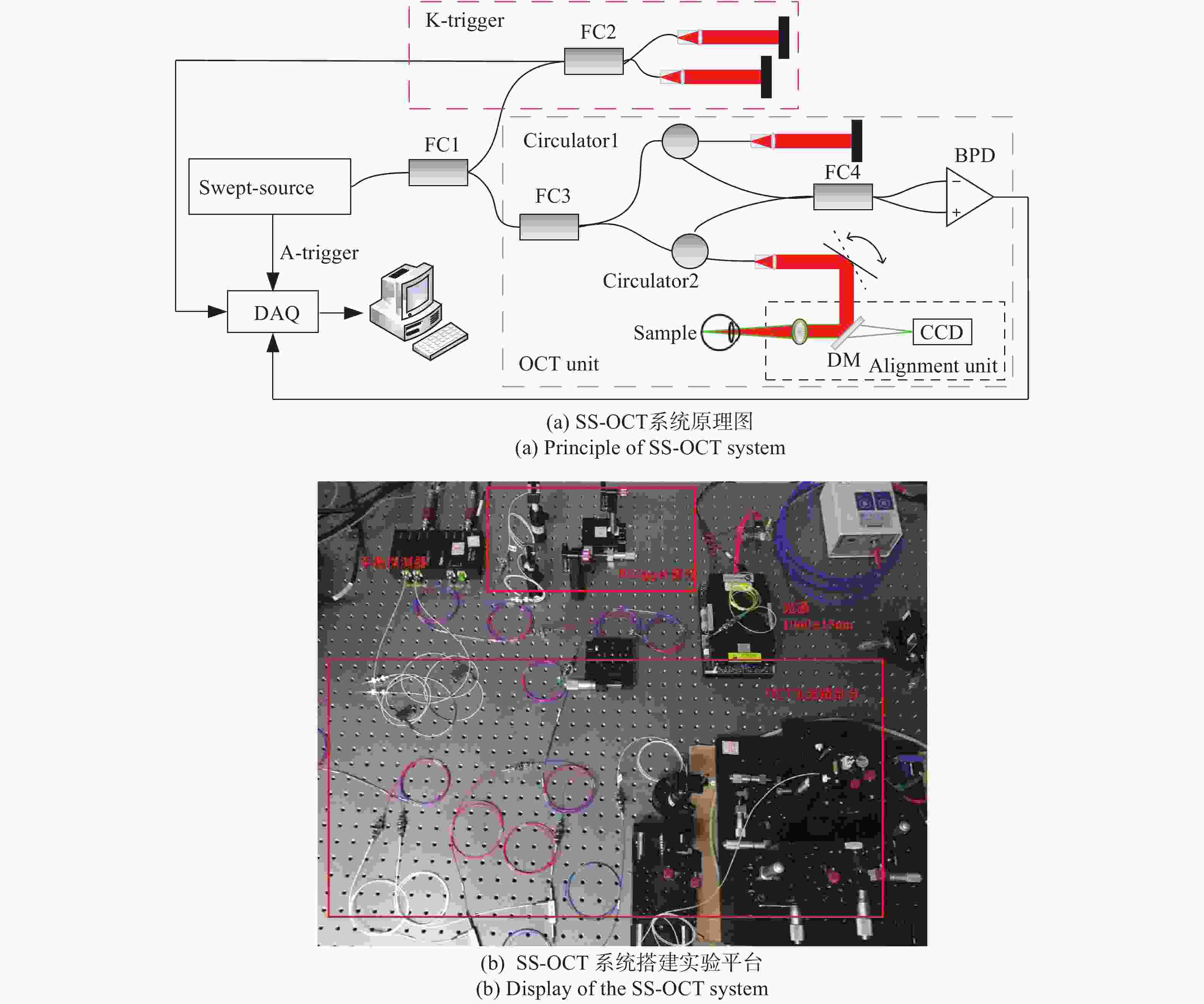
Figure 1. Fundamental principle and the display of SS-OCT system
系统分为OCT主光路部分,K-trigger重采样部分和角膜定位部分。随时间编码的宽带光源经耦合器分别输入到K-trigger标定光路和OCT主光路中。主光路样品臂光束依次经过角膜定位装置和扫描振镜入射到人眼,实现OCT的二维扫描,角膜定位装置使用可见光照明,反射光经二向色镜后被CCD收集;样品的后向散射光原路返回,与参考镜的反射光发生干涉后经平衡探测器转化为电信号进入采集卡;K-trigger标定光路中光束经平面镜反射后形成干涉信号;A-trigger信号为扫频光源发出的与光源扫频周期相同的TTL信号,下降沿时触发数据采集卡对K-trigger与OCT信号进行采集,采集完成后由PCIe总线上传到PC端,对OCT信号进行重采样和傅里叶变换处理,最终实现眼轴及眼前节的大量程成像。
-
SS-OCT系统数据处理流程如图2所示。首先,对光电转换器采集到的干涉信号进行光谱整形和
$k$ 域重采样处理,然后对信号傅里叶变换后提取样品信息,经图像增强后生成最终图像。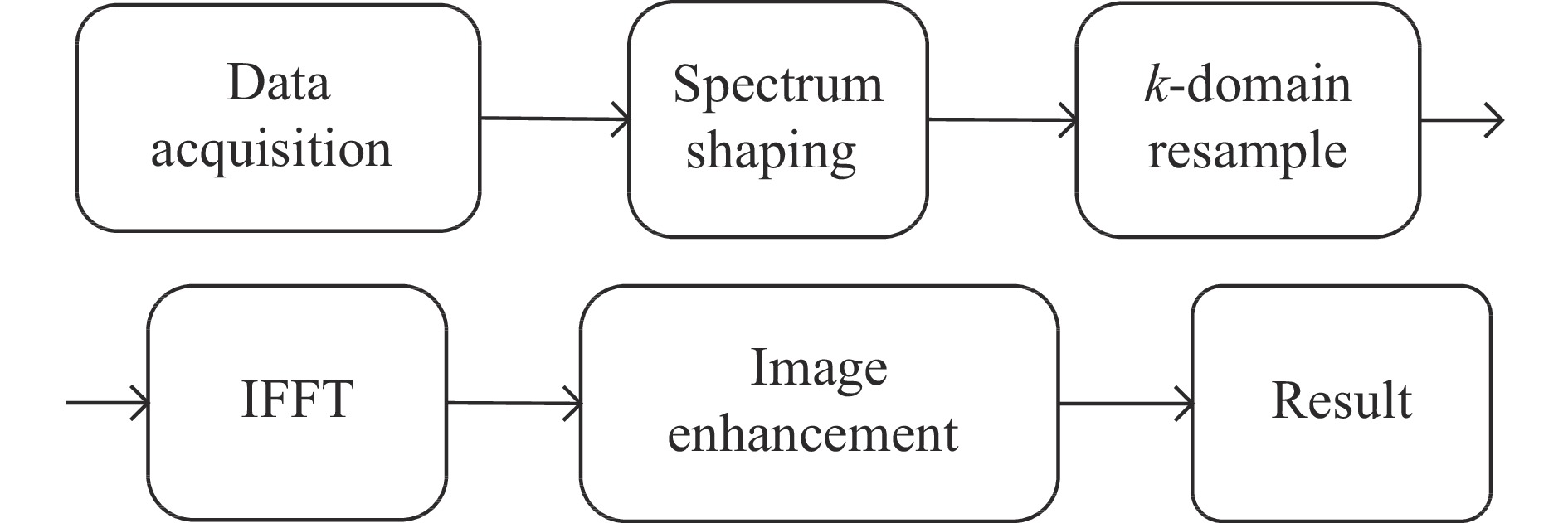
Figure 2. Flow chart of SS-OCT system data processing
-
SS-OCT系统由光源、采集卡、扫描振镜等多个模块构成,为实现模块之间的协调稳定运作,需利用扫频光源的A-trigger信号实现采集卡、扫描振镜等各组件之间的通信。图3为SS-OCT系统数据采集时序示意图。图中A-trigger是扫频光源触发信号,在单个扫频周期内对OCT以及K-trigger信号进行下降沿触发采样,再借由采集卡的辅助I/O口输出电平,控制信号发生器驱动扫描振镜工作,完成12 mm范围的横向扫描。
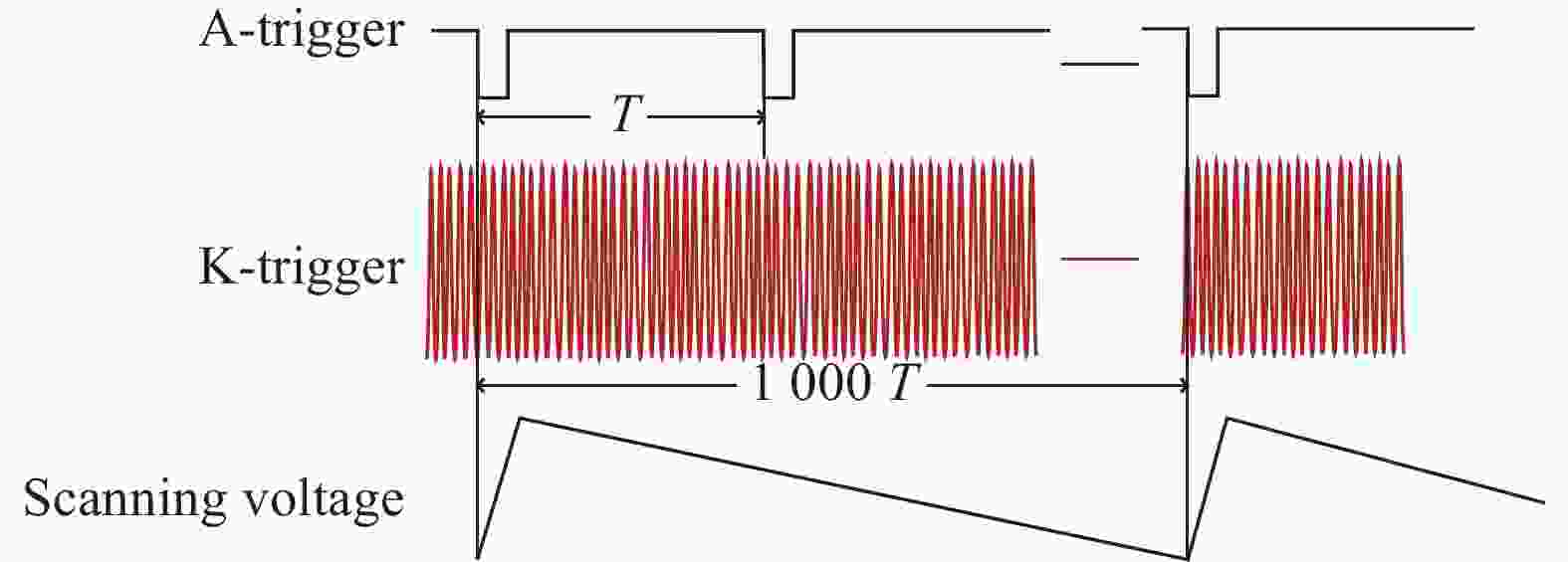
Figure 3. Schematic diagram of data acquisition sequence
-
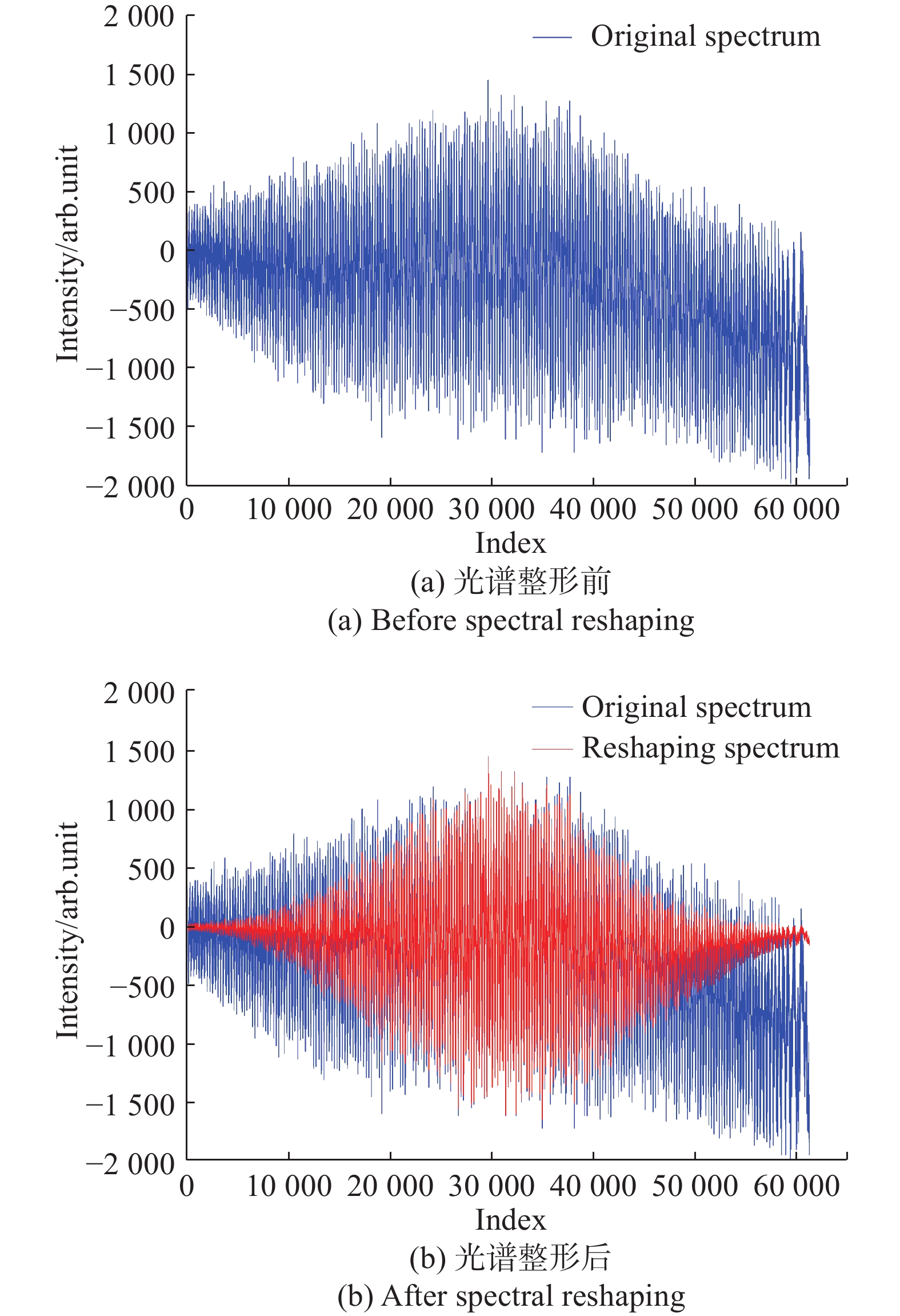
光源干涉光谱的包络形状会影响图像质量和系统分辨率。由于扫频光谱的分布并不是理想的高斯型,光谱曲线如图4(a)所示,对干涉信号进行傅里叶变换后得到的自相关函数存在旁瓣和脉冲展宽。为提高系统分辨率,采用基于窗口函数的光谱整形方法。实验表明,光谱整形后系统点扩散函数主瓣展宽、旁瓣减小。在实验中选取汉明窗(Hamming)的表达式为:

Figure 4. Sample's interference spectra before and after spectral reshaping
光谱整形之后的光谱
${S^{'}}\left( k \right)$ 与原始光谱$S\left( k \right)$ 的关系为:实验采用平面反射镜作为样品验证光谱整形效果,如图4所示,光谱整形前后干涉光谱包络有很大差异。图4(a)为光谱整形前的原始干涉光谱,光谱包络较大,峰值错位,呈现非高斯型;光谱整形后为规则的包络,信号展宽降低,如图4(b)所示。
-
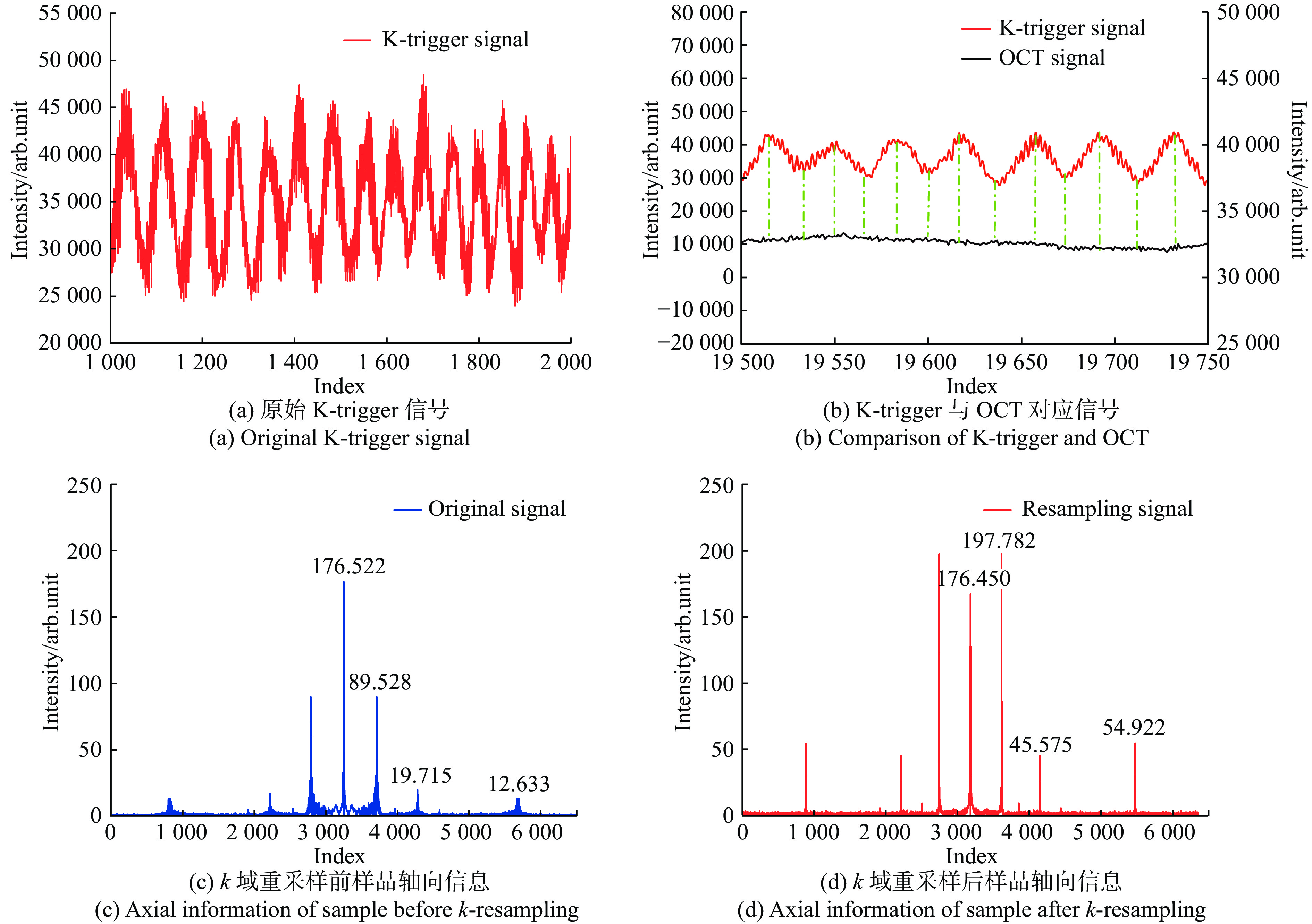
OCT信号的采样点数是决定系统干涉长度的重要依据[19],频域OCT系统中
$k$ 域重采样的方法主要有棱镜色散补偿[20]、电路K-clock时钟[21]、非均匀离散傅里叶变换[20]以及数值插值重采样[20]等。前两种均为硬件光谱重采样方法,系统复杂度高,而非均匀离散傅里叶变换不能采用快速傅里叶变换对数据进行处理,处理速度很低。因此文中选用最邻近数值插值的方法[22]对OCT信号进行$k$ 域重采样处理。光源产生的K-trigger信号和OCT信号如图5所示,受系统中背景噪声等影响,K-trigger信号为光程差恒定的类正弦信号,相邻峰值点间相位差均为π。考虑到系统中存在采样卡的硬件误差和背景噪声等因素,文中使用自适应峰值点提取与自适应误差校正相结合的算法,以实现对峰值点的提取[23]。通过实验可知,扫频光源的扫频频率及采集卡的采样率决定了K-trigger干涉信号稳态成分的相邻峰值点坐标差的理论平均值为
$S$ ,理论波动范围$\left[ {S - 15,S + 15} \right]$ ,为精确提取出K-trigger信号的峰值点,首先采用1×5平滑滤波器对信号进行平滑滤波,降低系统噪声的干扰;设平滑后的任一数据点强度为$f$ ,通过连续后向差分值$\Delta {f_n} = \left( {{f_{n + 1}} - {f_n}} \right) - \left( {{f_n} - {f_{n - 1}}} \right)$ 的符号判断极值点,重复计算至相邻极值点间的坐标差值$\Delta \in \left[ {S - 15,S + 15} \right]$ ,将此结果作为自适应峰值点提取的结果。计算连续周期内峰值点坐标差平均值$mea{n_j} = \frac{{\sum\limits_{j - m}^{j + m} {dif{f_{loca{l_j}}}} }}{{16}}$ ,其中$dif{f_{loca{l_j}}} = {n_j} - {n_{j - 1}}$ 为后向差分计算初步提取的峰值点中相邻两峰值点间坐标差值。由统计学规律可知,误差点一般与有效峰值点的坐标较为接近且满足$dif{f_{loca{l_j}}} < \dfrac{1}{2}mea{n_j}$ ,据此删去满足规律的误差点,以保证信号相邻峰值间的相位间隔为π。
Figure 5. K-trigger signal and signal before and after k-resampling
由于K-trigger与OCT为同源光,因此提取K-trigger信号中相位间隔为π的峰值点即可提取OCT信号在波数k域的均匀采样点,完成k域重采样。将重采样后的信号进行反傅里叶变换,即可提取轴向的A-scan信息,实验表明,重采样后样品信号增强为原始信号的2倍。
-
针对OCT图像中存在大量散斑噪声的问题[24],文中提出基于各向异性滤波的OCT图像增强算法。SS-OCT系统单帧成像时间为0.3 ms,连续帧图像间仅含有微小的刚性变换,因此使用对噪声敏感度小的相位相关算法[25, 26]对多帧图像进行配准。对于配准后的图像采用多帧差分的叠加方法,针对图像边缘较弱的问题,采用基于均值滤波的各向异性滤波方法(MF-PM filtering)进行边缘增强。图像增强流程如图6所示。

Figure 6. Flow chart of image enhancement
对于配准后的连续
$n$ 帧图像,使用相邻帧图像差分构造出$\frac{n}{2}$ 幅噪声图,$n$ 帧叠加图与噪声图作差即可突出目标区域从而提高OCT图像的对比度,得到噪声相对离散的OCT图像。多帧差分叠加公式如下:式中:
$oct(i,j)$ 是叠加后图像;$or{i_k}(i,j)$ 为配准后未叠加的单张原始图像。经实验验证,当$n = 6$ 时在保证成像速度的同时取得较好叠加结果。针对叠加后图像中依然存在的散斑噪声,对各向异性滤波方法进行改进,使用基于均值滤波的各向异性滤波方法(MF-PM filtering)进行边缘增强,最终得到边缘清晰、噪声被明显抑制的OCT图像。
各向异性扩散(PM)模型[27]利用梯度算子辨别由噪声和边缘引起的图像梯度变化,用邻域加权平均去除由噪声引起的小梯度变化,同时保留由边缘引起的大梯度变化。针对各向异性滤波遇强噪声易失效且易在梯度变化率大的位置引起“阶梯效应”,文中对各向异性滤波方法改进,基于均值滤波对散斑噪声处理的优势,用经过均值滤波平滑后的梯度模代替原始图像的梯度模,从而控制扩散的过程。
扩散系数
式中:
${I_0}$ 为原图像;$\left\| {\nabla I} \right\|$ 可视为边缘检测器;${M_\sigma }$ 为中值滤波窗口;$k$ 为扩散门限。 -
根据第二节中介绍的系统原理的搭建实验平台如图1(b)所示。文中使用中心波长1060 nm,带宽30 nm的定制扫频光源(AXSUN-1060),光源频率3 kHz相干长度为69 mm,光源输出最大功率30 mW。采用外部搭建的重采样K-trigger标定光路对OCT信号进行重构,实现眼轴及眼前节的实时成像。
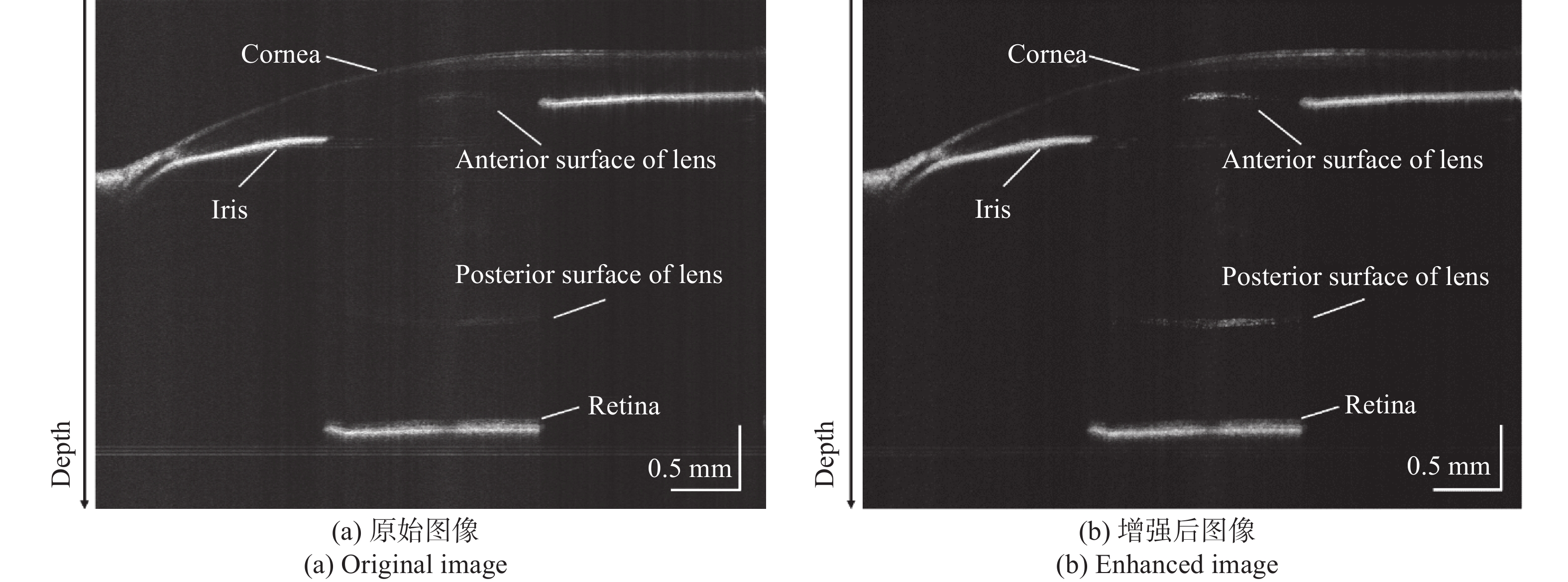
该系统使用离体仍具生理活性的鱼眼进行实验,得到图7(a)所示的原始图像,实现生物眼的眼轴及眼前节的大量程成像。对图7(a)按照上节方法进行增强处理后得到信号明显增强、噪声大幅度抑制且边缘轮廓清晰的图像,如图7(b)所示。角膜表面、晶状体前后表面及眼底各层结构均清晰可见。
由于实验中所用离体生物眼的各层折射率与相对位置未知,因此对等比例人眼模型(Ocular,OEMI-7)成像,验证该系统测量结果的准确性。
为解决系统因扩大干涉范围所引起的纵向分辨率降低、减小扫描过程中由于扫描振镜机械振动造成纵向伪影和展宽的问题,实验中控制扫描振镜在0.1 cm的横向范围内进行扫描,以眼轴位置为中心,前后各取5组A-scan信号,生成如图8所示的二维结果,通过各层数据均值作为各层结构的实际位置对实验结果进行校准。图8中各层信号从左至右分别对应的眼球内部结构依次为角膜前、后表面,晶状体前、后表面以及眼底。

Figure 7. Original image and the enhanced results

Figure 8. Full-scale eye model measurement result
文中将实验所得数据点数依据眼内各组织折射率折算为相应生物学尺寸,从而得到各层组织参数。已知空气中光程差改变所对应数据点数的变化
${R_{air}}$ ,根据人眼各层结构所对应的点数${D_{air}}$ 与折射率${n_i}$ 可知各结构的实际尺寸${D_n} = {D_{air}}/\left( {{R_{air}} \cdot {n_i}} \right)$ ,人眼内角膜、前房、晶状体、玻璃体及视网膜的折射率分别为1.387、 1.342、1.415、 1.341、 1.380。根据参数计算出样品各层结构尺寸如表1所示。Measurement CCT/mm AD/mm LT/mm VT/mm AL/mm Sample size 0.33 3.06 4.06 18.97 26.42 Result without calibration 0.35 3.08 4.07 19.10 26.60 Result after calibration 0.32 3.07 4.07 19.00 26.46 Error value 0.01 0.01 0.01 0.03 0.04 Table 1. Comparison between the measurement of the parameters of each layer of the eye axis and the actual sample size
该系统通过对多组A-scan数据求取均值的方法校准可精确得出等比例眼模型眼轴的各层参数,表1中分别展示了校准前后的各结构测量尺寸。与模型实际尺寸进行对比可知,本系统对角膜厚度(CCT)、前房深度(AD)、晶状体厚度(LT)、玻璃体厚度(VT)等组织的测量误差分别为0.01 mm、0.01 mm、0.01 mm和0.03 mm,眼轴测量误差为0.04 mm,测量重复性满足临床医学成像的要求。
参考文献[13]使用可变光程的参考臂并拼接成像,系统的测量误差和实时性均低于该系统;参考文献[14]中双波段双聚焦OCT在三维成像方面取得了较好的效果,但成像误差为0.77 mm,成像时间10 s,远超过该系统提出的方法;参考文献[28]方法误差为0.02 mm,但耗时较长。通过表2中各分段检测方法对比可知,该系统可以稳定实现对生物眼轴的实时精确测量,各层结构平均测量误差小于0.02 mm,眼轴平均测量误差0.04 mm,成像时间0.45 s,兼具测量误差小与成像速度快的优势,在无需对各结构进行长度拼接的情况下,成像范围可覆盖全眼。
Source Method Measurement
error/mmImaging dimension Number of
layersTime/s Tao[13] Reference arm with variable optical path 0.45 2D 5 0.9 Shanghai Jiao Tong University[14] Dual band dual focus OCT 0.77 2D/3D 5 10 Marco[28] Reference arm with variable optical path 0.02 2D 5 3.4-4 This system Large-scale measuring system 0.04 2D 5 0.45 Table 2. Comparison results with traditional segmented measurement system
-
根据眼轴测量及眼前节成像的大量程测量深度和高分辨率需求,设计了一种基于SS-OCT的大量程眼轴及眼前节一体化成像系统。通过外部搭建的K-trigger标定光路提高采样点数、并通过k域重采样和光谱整形提高图像质量和系统分辨率;提出基于各向异性的保边去噪算法对图像进行增强,实现了正常眼眼轴范围的实时测量以及眼前节范围内的清晰成像。横向成像范围12 mm,平均测量误差小于0.02 mm,眼轴测量误差0.04 mm,成像时间0.45 s。目前该系统尚未对不健康眼的样本进行实验,系统生物成像的鲁棒性有待一进步提高。
Integrated imaging system of eye axis and anterior segment based on SS-OCT
doi: 10.3788/IRLA202049.0413006
- Received Date: 2019-12-11
- Rev Recd Date: 2020-01-24
- Publish Date: 2020-04-24
-
Key words:
- swept-source optical coherence tomography /
- eye axis measurement /
- anterior imaging /
- k
-domain resampling / - image enhancement
Abstract: A system of Swept-Source Optical Coherence Tomography for synchronous measurement of the eye axis with large imaging depth range and anterior segments was proposed to solve the problem that traditional ophthalmic axis measuring instruments can not achieve the functions of large imaging depth in axial measurement and imaging on anterior simultaneously. The proposed system increased the imaging depth by designing a infrared swept source with wide range. Then a calibrated optical path was built for k-domain resampling to improve the imaging quality in large range interference. Thus the high precision axial information and the anterior segment image could be obtained simultaneously. To solve the problem of low SNR of OCT images in this system, an edge-preserving denoising algorithm based on anisotropy filtering was proposed to suppress the speckle noise, which effectively improved the image contrast. Using the equal-proportion human eye model and vitro fish eye imaging respectively, the experimental result shows that the imaging depth of the system can reach 69 mm. The 12 mm anterior segment can be completely scanned in transverse direction, while the axial five-layer structure is accurately measured. The average measurement error of axial length is 0.04 mm, and the imaging time is about 0.45 s, which fulfil the real-time requirements of clinical medical application.


 DownLoad: